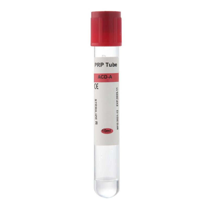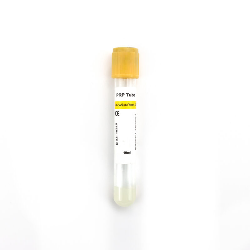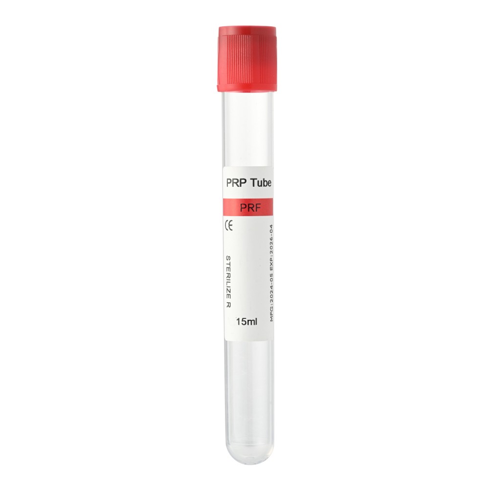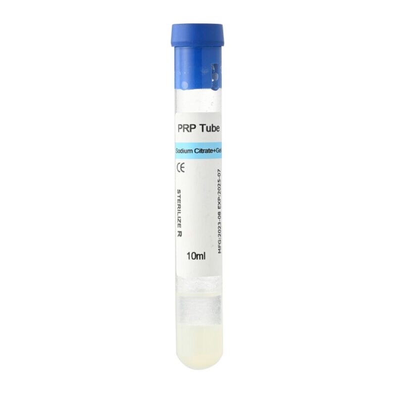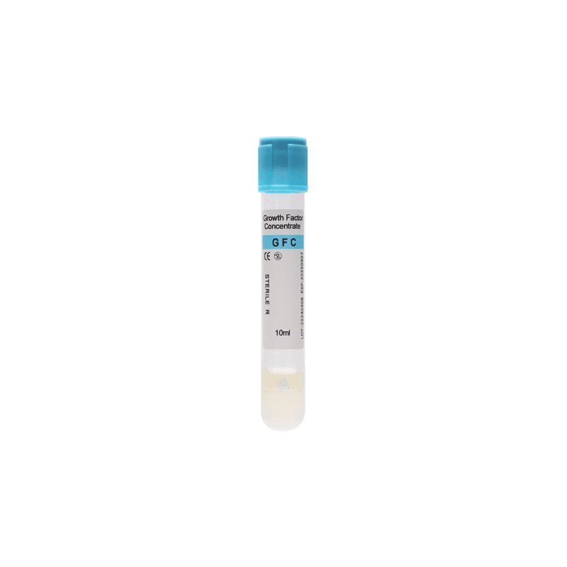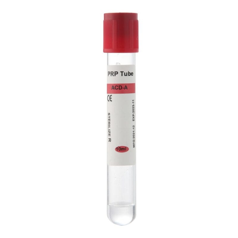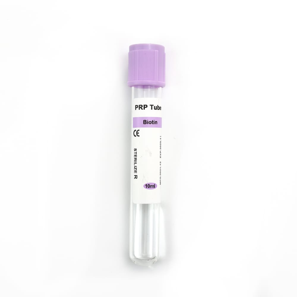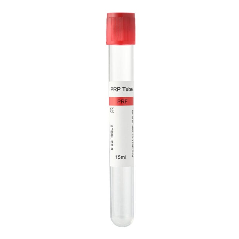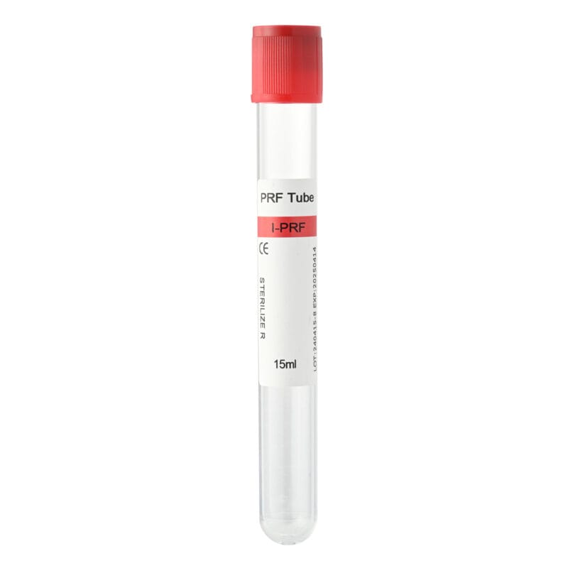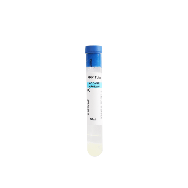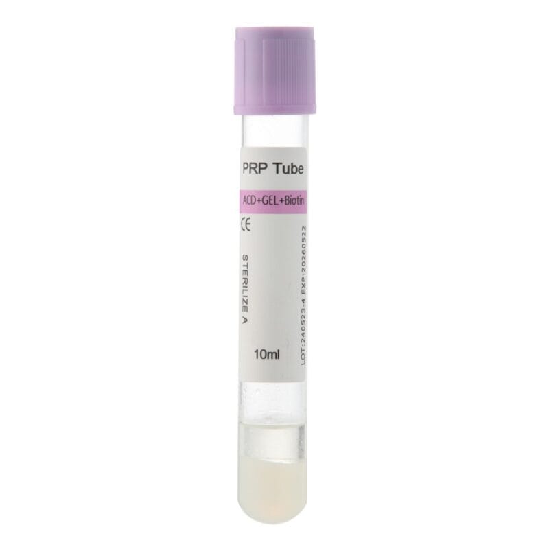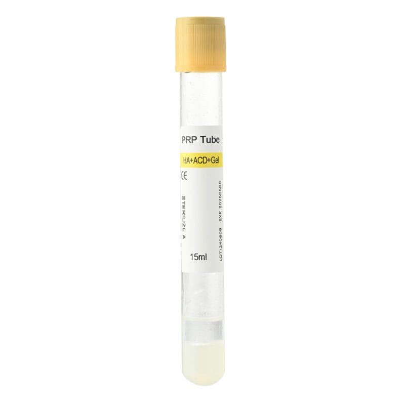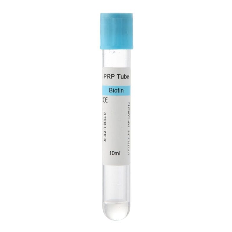
Sticky Bone in Dentistry is a bone grafting technique that combines particulate bone graft materials with a fibrin-based preparation derived from the patient’s blood. The result is a cohesive, moldable graft that dentists can shape and place in areas requiring bone regeneration.
Unlike conventional loose bone graft particles, sticky bone forms a connected mass that is easier to handle during surgery. Its fibrin network helps hold graft particles together, while the bone substitute provides a framework for new bone formation.
This technique has gained attention in dental implantology, periodontal surgery, and alveolar bone augmentation. Injectable platelet-rich fibrin (i-PRF) is particularly relevant because its liquid form can be mixed with graft particles before fibrin polymerization occurs.
However, successful preparation requires appropriate blood collection tubes, centrifugation conditions, and surgical techniques. Understanding these factors helps dental professionals select suitable materials for their regenerative procedures.
This guide explains how sticky bone works, how it is prepared, its main dental applications, and what clinics should consider when choosing PRF tubes.
What Is Sticky Bone in Dentistry and How Does It Work?
Sticky bone is a composite graft material in which bone particles are incorporated into a fibrin network. It is also associated with techniques described as mineralized plasmatic matrix or fibrin-assisted bone grafting.
The main purpose is to improve graft cohesion while combining the structural properties of bone substitutes with the biological components of autologous blood preparations.
The Main Components of Sticky Bone
Three elements contribute to the characteristics of the final graft.
Bone Graft Material
Particulate bone substitutes provide an osteoconductive framework that supports new bone formation.
Common grafting materials include:
- Autografts: Bone collected from the patient’s own body.
- Allografts: Human donor-derived bone materials.
- Xenografts: Bone substitutes obtained from another species.
- Alloplasts: Synthetic substitutes, including selected calcium phosphate materials.
Each material has different characteristics related to resorption, volume maintenance, and biological performance. Dentists select the graft according to the defect and treatment objective.
Platelet-Rich Fibrin (PRF)
PRF is an autologous blood-derived preparation containing a fibrin matrix, platelets, and varying amounts of leukocytes and other cellular components.
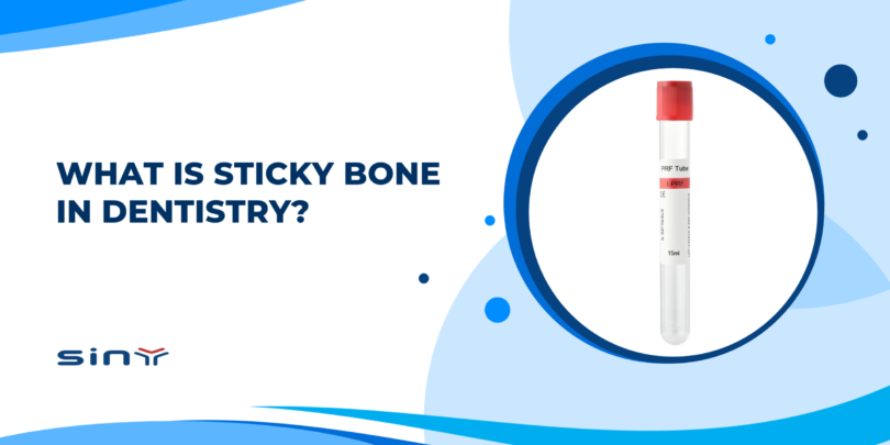
It is produced through centrifugation using an appropriate blood collection system. Depending on the preparation protocol, PRF may be obtained as a solid clot, membrane, or injectable liquid.
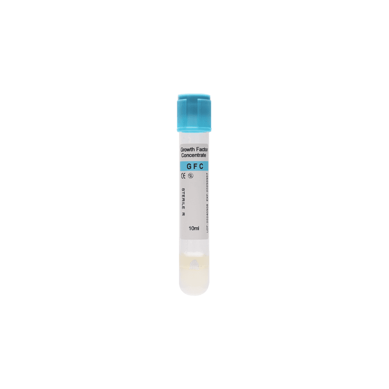
The liquid form, known as i-PRF, is commonly used for sticky bone preparation.
Fibrin Network
Fibrin provides the connection between the individual graft particles.
As coagulation occurs, fibrinogen converts into fibrin. The resulting network surrounds the bone particles and gradually creates a cohesive structure.
This helps clinicians manipulate the graft without excessive particle scattering.
How Does Sticky Bone Support Bone Regeneration?
The bone substitute and fibrin preparation perform different but complementary roles.
Bone particles provide structural support and a surface for new bone formation. Meanwhile, the fibrin matrix contributes to graft cohesion and creates a temporary environment for cellular activity.
PRF also contains signaling molecules associated with tissue repair, including platelet-derived growth factor (PDGF) and transforming growth factor beta (TGF-β).
These components may support processes involved in healing. However, their presence does not guarantee faster bone regeneration or better clinical outcomes in every patient.
The final result depends on the graft material, defect characteristics, preparation protocol, and surgical management.
How Is Sticky Bone Prepared Using PRF?
Sticky bone can be prepared using several fibrin-based methods. Two approaches involve liquid i-PRF or components obtained from conventional PRF clots.
The i-PRF method is particularly useful because liquid fibrin can distribute throughout particulate graft material before coagulation.
Step 1: Blood Collection
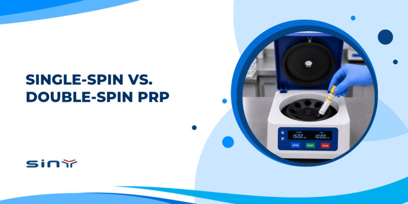
The procedure begins with collecting the patient’s venous blood into tubes compatible with the selected PRF protocol.
Conventional PRF and i-PRF techniques based on natural coagulation generally use tubes without anticoagulants.
Tube material and surface characteristics can influence coagulation. Therefore, clinicians should select products designed for their intended preparation method.
Blood collection and processing must follow appropriate clinical and aseptic procedures.
Step 2: Centrifugation
The collected blood undergoes centrifugation to separate its components.
For conventional PRF, the process produces a solid fibrin clot that may be compressed into a membrane.
For i-PRF, a suitable protocol produces a liquid fraction that can be collected before substantial fibrin polymerization.
Centrifugation conditions vary with the equipment and preparation system. Relative centrifugal force (RCF), rotor geometry, processing time, and tube characteristics all matter.
Consequently, RPM settings from one centrifuge should not be applied directly to another without checking the corresponding RCF.
Step 3: Mixing With Bone Graft Material
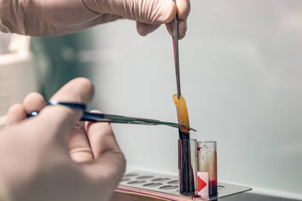
The clinician combines the prepared liquid fibrin fraction with particulate bone graft material in a sterile container.
As coagulation progresses, the fibrin network develops around the particles and gradually binds them together.
The mixture becomes more cohesive and can be shaped to fit the intended bone defect.
Some alternative methods incorporate fragmented conventional PRF clots, PRF exudate, or other autologous fibrin preparations.
Step 4: Graft Placement
Once the mixture reaches the desired consistency, the clinician positions it in the prepared defect.
Its cohesive structure can improve handling and help reduce particle displacement during placement.
However, additional stabilization or barrier membranes may still be necessary, depending on the defect and surgical technique.
Conventional PRF vs. i-PRF for Sticky Bone
| Feature | Conventional PRF | Injectable PRF (i-PRF) |
|---|---|---|
| Typical form | Solid fibrin clot | Liquid before polymerization |
| Preparation objective | Obtain a fibrin clot or membrane | Obtain a usable liquid fibrin preparation |
| Role in bone grafting | Membrane coverage or incorporation into graft mixtures | Mixing directly with particulate graft material |
| Handling characteristics | Can be trimmed, compressed, or fragmented | Distributes through graft particles before clotting |
| Common applications | Soft-tissue management and regenerative procedures | Sticky bone and selected regenerative applications |
Both preparations have roles in regenerative dentistry. However, different collection tubes and processing protocols may be required.
Applications of Sticky Bone in Dental Procedures
Dental bone grafting is commonly performed when existing bone is insufficient for a planned treatment or requires reconstruction.
Sticky Bone in Dentistry is used in several regenerative procedures where graft cohesion and adaptation to the surgical site are important.
Dental Implant Placement
Dental implants require adequate bone volume and suitable surrounding tissue conditions.
When the available bone is insufficient, clinicians may perform bone augmentation before or during implant placement.
A cohesive graft mixture can be shaped around selected implant-related defects, making particulate material easier to position.
However, sticky bone does not replace proper implant planning, surgical stability, or suitable graft containment.
Alveolar Ridge Augmentation
The alveolar ridge supports natural teeth and provides the foundation for dental implants.
Following tooth loss, bone remodeling may reduce ridge height or width. This can complicate future restoration.
Ridge augmentation aims to restore the dimensions needed for treatment.
A fibrin-bound graft can adapt to certain irregular defects and help maintain the particulate material as a connected mass during placement.
Depending on the defect, barrier membranes or fixation techniques may still be needed for space maintenance.
Socket Preservation After Extraction
Tooth extraction commonly leads to changes in alveolar ridge dimensions.
Socket preservation procedures aim to reduce these changes and maintain bone volume for future restorative treatment.
Dentists may use particulate graft materials to fill selected extraction sockets.
When combined with a fibrin preparation, the particles become easier to handle and adapt to the socket.
However, not every extraction requires grafting, and sticky bone is not necessary in every socket preservation procedure.
Sinus Floor Augmentation
Insufficient bone height in the posterior upper jaw can make dental implant placement challenging.
Sinus floor augmentation creates additional space for bone grafting beneath the maxillary sinus membrane.
A cohesive graft may simplify material handling during selected sinus augmentation procedures.
Nevertheless, membrane integrity, graft stability, available bone height, and surgical technique remain essential considerations.
Periodontal Bone Defects
Advanced periodontal disease can damage the bone supporting natural teeth.
Certain intrabony defects may be treated using regenerative techniques involving bone substitutes and biologically active materials.
Sticky bone has been investigated in these procedures because its fibrin structure may improve graft handling and support the local healing environment.
However, periodontal treatment outcomes depend on infection control, defect anatomy, oral hygiene, and patient-related factors.
Long-term evidence is still needed to establish the additional clinical benefits of individual sticky bone protocols.
Benefits and Limitations of Sticky Bone Grafts
The main practical advantage of sticky bone is its cohesive structure. This can improve graft handling while incorporating biological components derived from the patient’s blood.
However, these characteristics should not be confused with guaranteed improvements in clinical outcomes.
Potential Benefits
Improved Graft Cohesion
Fibrin helps connect particulate graft material into a moldable mass.
This reduces particle scattering during manipulation and can make placement easier than handling loose granules.
Adaptation to Irregular Defects
Bone defects vary in shape and size.
A cohesive graft can be molded to fit selected defects, which may be useful during ridge augmentation and implant-related procedures.
Autologous Biological Components
PRF contains fibrin, platelets, and associated signaling molecules derived from the patient’s blood.
These components participate in tissue repair and may contribute to the regenerative environment.
However, the bone substitute itself may still be donor-derived, animal-derived, or synthetic.
Potential Healing Support
The fibrin matrix provides a temporary scaffold for cellular activity.
Some studies report favorable healing or regenerative findings. Nevertheless, the strength of evidence varies between procedures and preparation methods.
Limitations and Clinical Considerations
Variable Clinical Evidence
Although sticky bone has demonstrated promising handling characteristics, clinical studies use different graft materials, centrifugation protocols, and outcome measures.
More standardized research is needed to determine whether specific techniques consistently improve long-term bone regeneration.
Preparation Sensitivity
Blood collection, tube selection, centrifugation, and handling can affect the final fibrin preparation.
Inconsistent processing may produce different material characteristics.
Time-Sensitive Handling
Liquid i-PRF gradually polymerizes after collection.
Delayed processing may reduce the time available to mix it evenly with graft particles.
Additional Stabilization May Be Necessary
A cohesive graft does not automatically provide sufficient space maintenance.
Certain defects require membranes, fixation, or other stabilization techniques.
For these reasons, dentists should select the technique according to the clinical indication rather than assuming it is superior to conventional bone grafting.
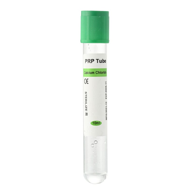
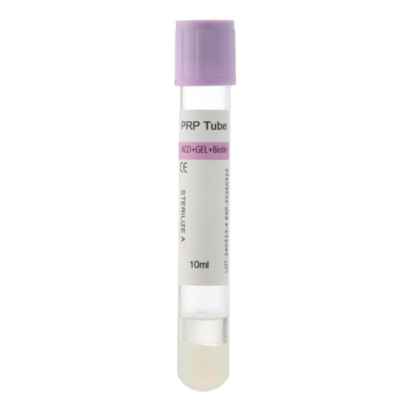
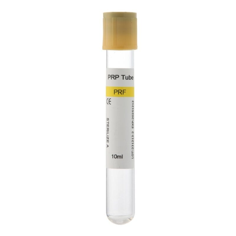
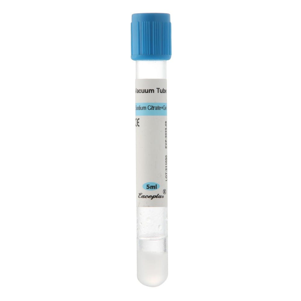
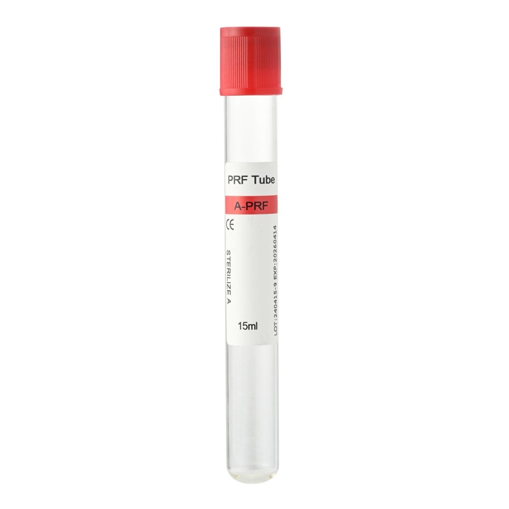
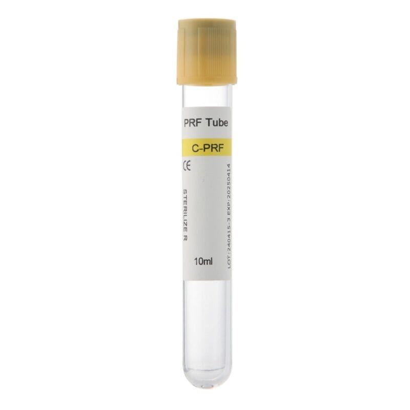
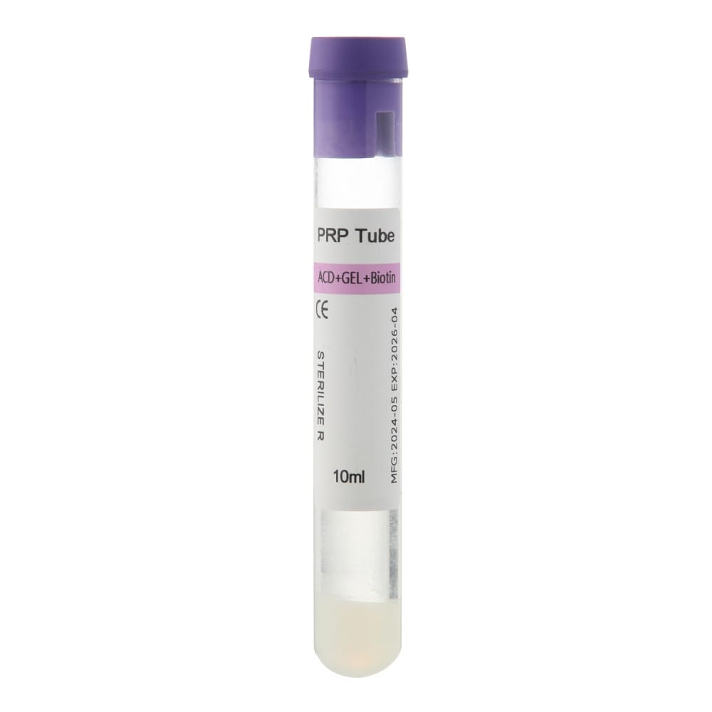
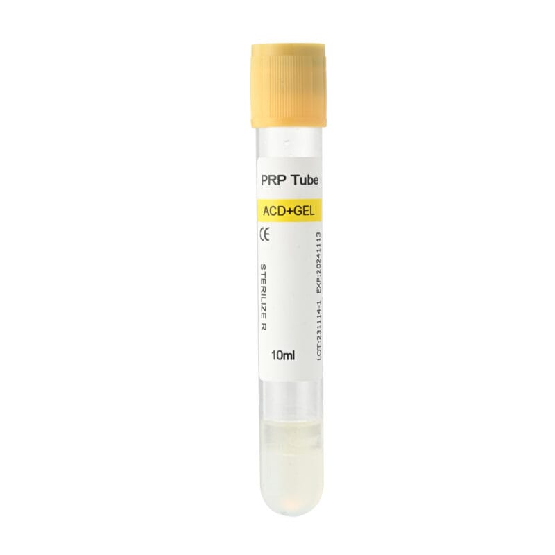
How to Choose PRF Tubes for Sticky Bone Preparation
Selecting suitable blood collection tubes is an important part of PRF preparation.
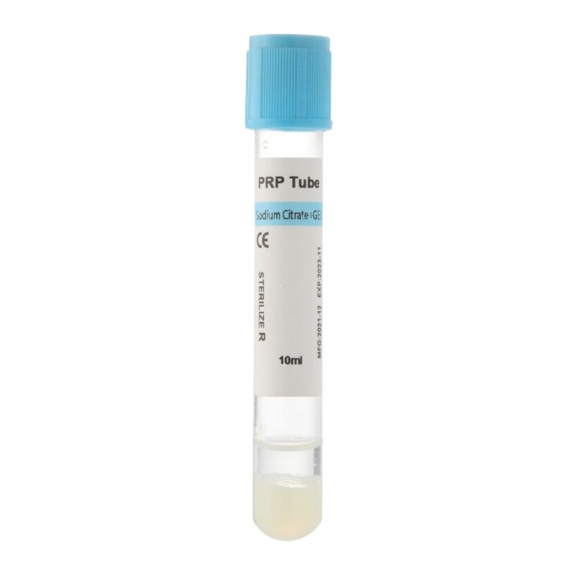
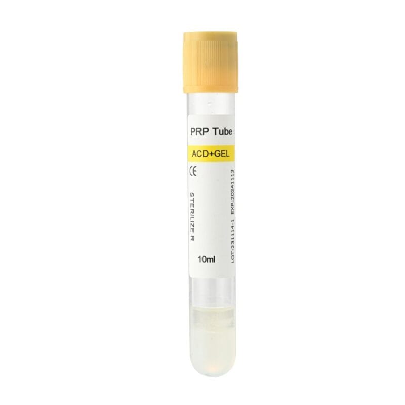
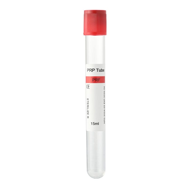
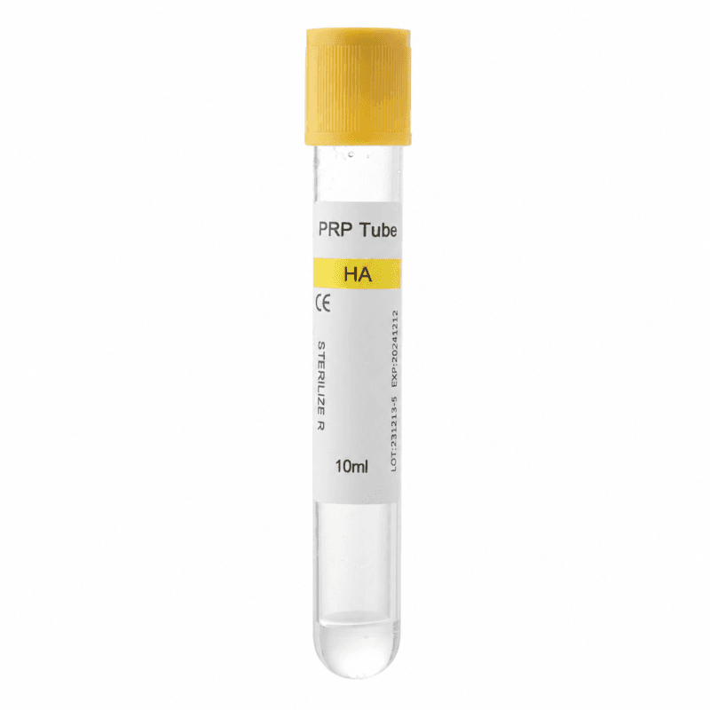
For dental clinics and distributors, the decision should consider the required fibrin form, tube material, surface characteristics, additives, and centrifuge compatibility.
Not every tube labeled as a PRF tube is suitable for every preparation method.
Conventional PRF Tubes vs. i-PRF Tubes
Clinics should first determine whether their procedure requires solid PRF or injectable fibrin.
Conventional PRF Tubes
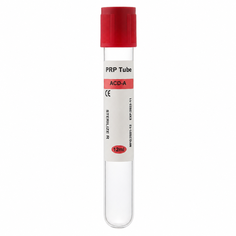
These tubes are intended for suitable protocols that produce solid fibrin clots.
The resulting material may be compressed into membranes or incorporated into selected grafting techniques.
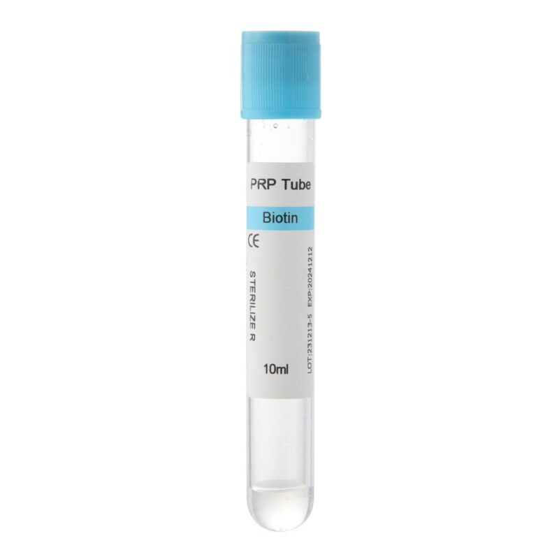
i-PRF Tubes
These tubes are intended for protocols that obtain a liquid fibrin preparation before substantial clot formation.
Liquid i-PRF can be mixed with particulate bone substitutes to produce a cohesive graft.
Although both preparations involve blood centrifugation, their collection and processing requirements may differ.
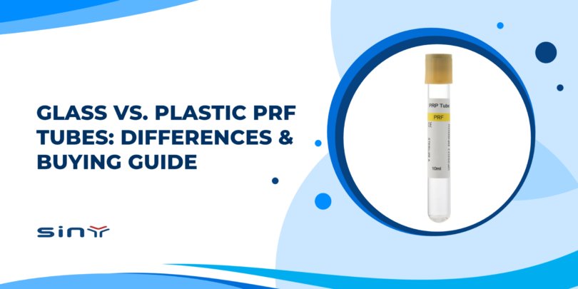
Glass vs. Plastic PRF Tubes
Tube material can influence coagulation behavior and the characteristics of the collected fibrin preparation.
Glass Tubes
Glass surfaces can promote contact activation of the coagulation process.
For this reason, suitable glass collection tubes are commonly associated with conventional solid PRF preparation.
However, tube suitability also depends on surface treatment, manufacturing characteristics, and the specific preparation protocol.
Plastic Tubes
Plastic collection tubes may have different coagulation characteristics because their surfaces interact differently with blood.
Certain additive-free, appropriately designed plastic tubes are used in liquid i-PRF protocols.
However, not all plastic tubes behave identically. Surface coatings, additives, and processing conditions can change their performance.
The material alone does not determine whether a tube is appropriate for conventional PRF or i-PRF.
Why Additives and Surface Coatings Matter
For PRF techniques based on natural coagulation, anticoagulants such as EDTA, citrate, and heparin are generally unsuitable because they interfere with the clotting process.
However, an absence of anticoagulants does not necessarily mean that a tube is free from other additives or surface treatments.
Some blood collection tubes contain silica-based clot activators or silicone-related coatings that can affect coagulation.
Certain research has raised concerns about possible particle contamination from specific clot-activating tube systems used for PRF preparation.
Therefore, dental clinics should check the manufacturer’s product information rather than assuming that every plain-looking collection tube is additive-free.
The key consideration is whether the complete collection system is appropriate for the intended clinical preparation.
Check Centrifuge Compatibility
A suitable PRF tube must also be compatible with the clinic’s centrifuge.
Important factors include tube dimensions, collection volume, rotor compatibility, and the required centrifugation protocol.
RCF is particularly important because identical RPM values can generate different centrifugal forces in centrifuges with different rotor radii.
For consistent preparation, clinics should use the tube and centrifuge combination specified by their selected protocol.
PRF Tube Purchasing Checklist
| Buying Consideration | What Buyers Should Check | Why It Matters |
|---|---|---|
| PRF preparation type | Conventional PRF or liquid i-PRF | Determines the intended tube application |
| Tube material | Glass or plastic, including surface properties | May influence coagulation behavior |
| Additives and coatings | Anticoagulants, clot activators, and surface treatments | Can affect fibrin formation and preparation quality |
| Centrifuge compatibility | Tube dimensions, rotor fit, and RCF requirements | Supports consistent processing |
| Sterility and intended use | Sterile packaging and manufacturer instructions | Important for clinical blood processing |
| Product consistency | Specifications, labeling, and supply reliability | Helps clinics maintain standardized workflows |
Consider Long-Term Purchasing Requirements
Clinics using both conventional PRF and i-PRF may need separate tube types for different procedures.
Distributors should also confirm product specifications, intended applications, packaging options, and availability before making bulk purchasing decisions.
A consistent supply of appropriate collection tubes can help clinics standardize their preparation workflow.
Ultimately, buyers should prioritize tube suitability and process compatibility rather than selecting products based only on price, appearance, or material.
Frequently Asked Questions About Sticky Bone in Dentistry
1. What is sticky bone made of?
Sticky bone is typically made by combining particulate bone graft material with a fibrin-based preparation derived from the patient’s blood. Injectable PRF or autologous fibrin glue is commonly used to create the cohesive structure that connects the graft particles.
2. Is sticky bone the same as PRF?
No. PRF is a blood-derived fibrin preparation, while sticky bone combines a fibrin preparation with particulate bone graft material. PRF contributes the fibrin network, while the bone substitute provides the structural framework for bone regeneration.
3. Why is i-PRF used for sticky bone?
i-PRF remains liquid for a limited period after preparation. This allows it to mix with bone graft particles before coagulation creates a cohesive fibrin network. Its handling properties make it useful for selected bone augmentation procedures.
4. Can conventional PRF tubes be used to prepare sticky bone?
Conventional PRF can be incorporated into certain sticky bone techniques, including methods using fragmented fibrin clots or PRF exudate. However, producing liquid i-PRF requires a compatible tube and preparation protocol. Not every conventional PRF tube is suitable for liquid fibrin preparation.
5. Does sticky bone improve dental implant success rates?
Sticky bone may improve graft handling and stability in selected procedures. However, current evidence does not establish that it consistently increases dental implant success rates compared with conventional grafting. Outcomes depend on surgical technique, patient condition, and bone quality.
6. How long does sticky bone take to form?
Formation time varies according to the fibrin preparation, graft material, tube characteristics, and processing conditions. Clinicians should follow their selected preparation protocol rather than relying on a universal clotting time.
7. Is sticky bone better than traditional bone grafting?
Neither technique is automatically superior for every patient. Sticky bone offers handling advantages, but its additional regenerative benefits depend on the clinical indication and preparation method. Conventional bone grafting remains an established treatment option.
8. What should dental clinics consider when buying PRF tubes?
Clinics should evaluate the intended PRF type, tube material, additives, surface characteristics, collection volume, sterility, and centrifuge compatibility. Buyers should also confirm manufacturer instructions and whether the tube is suitable for their specific preparation protocol.
Final Thoughts
Sticky bone combines particulate bone substitutes with fibrin-based preparations to create a cohesive graft for selected dental regenerative procedures. Its moldable structure can simplify graft placement, while its biological components may support the healing environment. Nevertheless, predictable treatment still depends on appropriate case selection, careful surgical technique, and consistent preparation.
For dental clinics and medical distributors, choosing suitable blood collection tubes is an important part of the workflow. Siny PRP offers both conventional PRF tubes and i-PRF tubes for different platelet-rich fibrin preparation needs. Explore the available products at Siny PRP or contact our team to discuss PRF tube options for your dental clinic or distribution business.