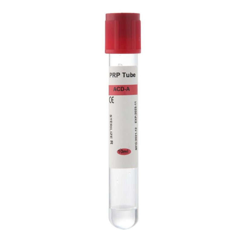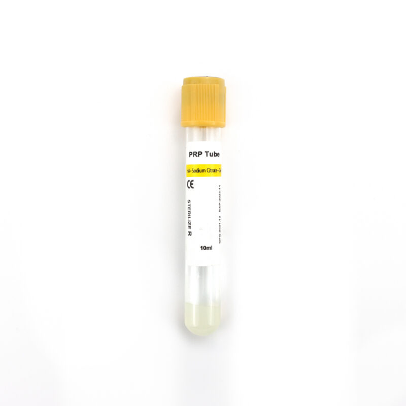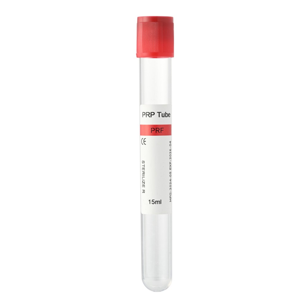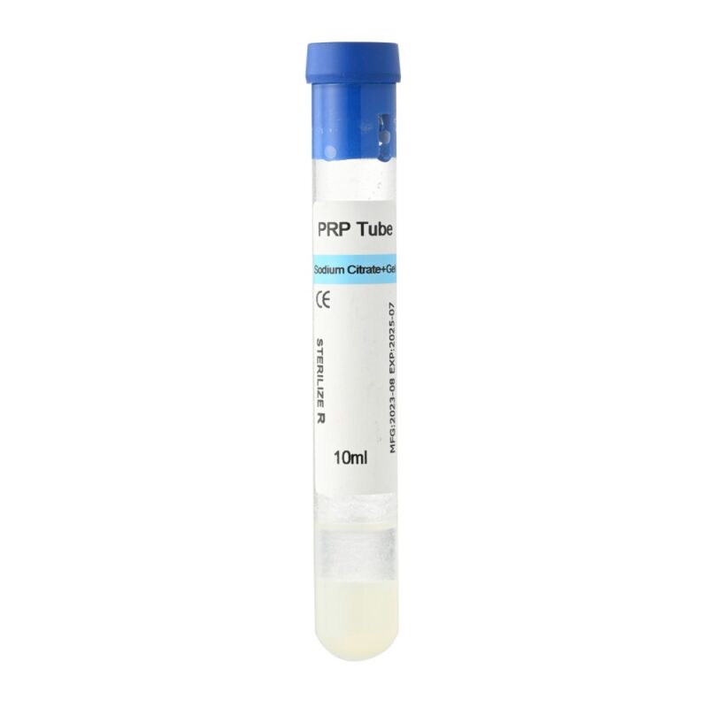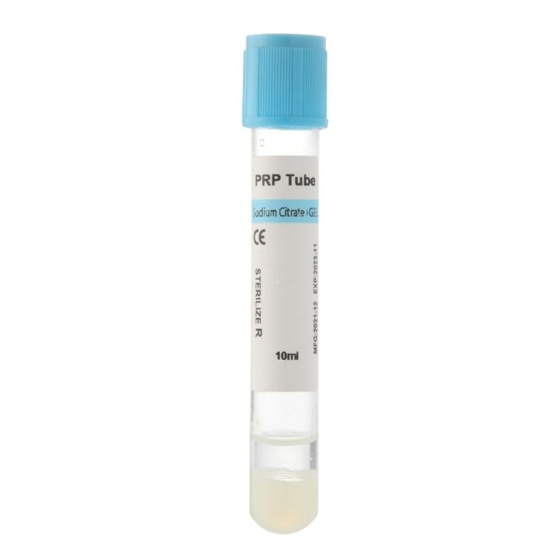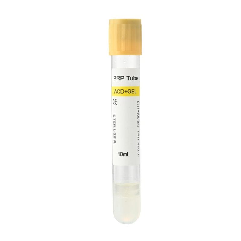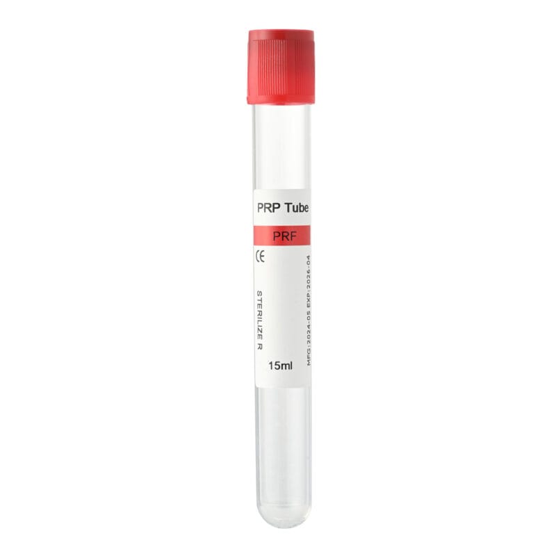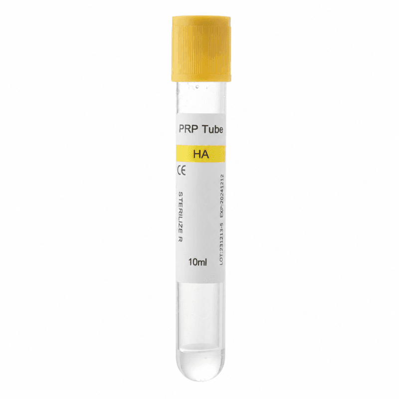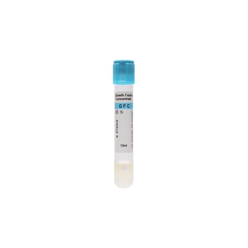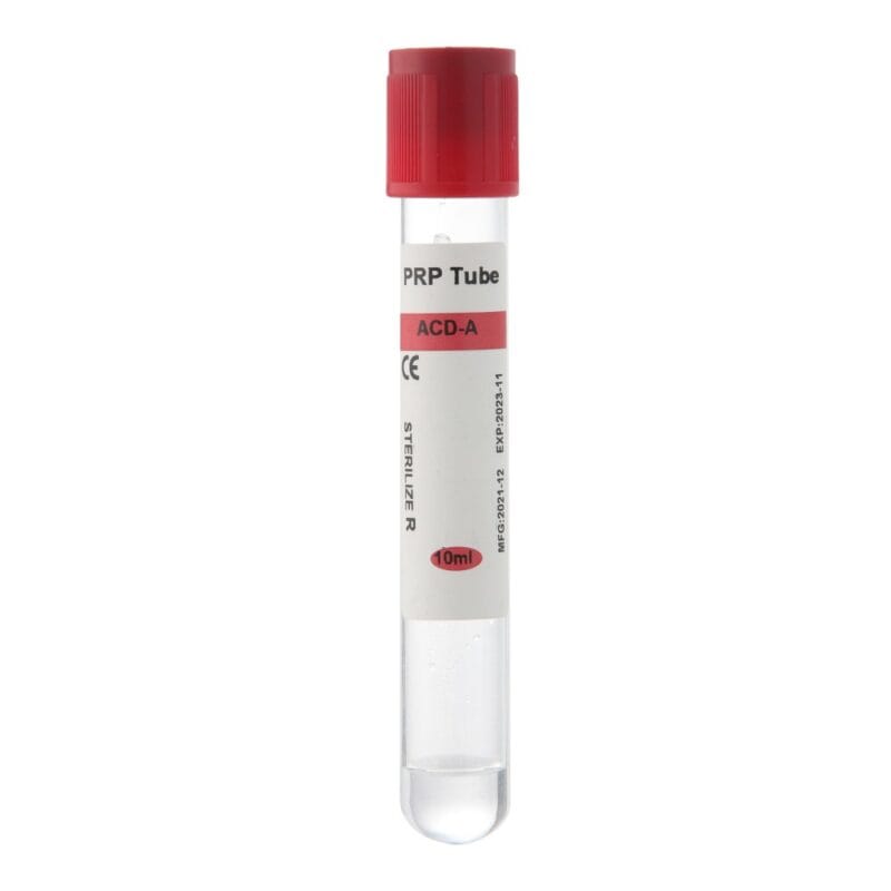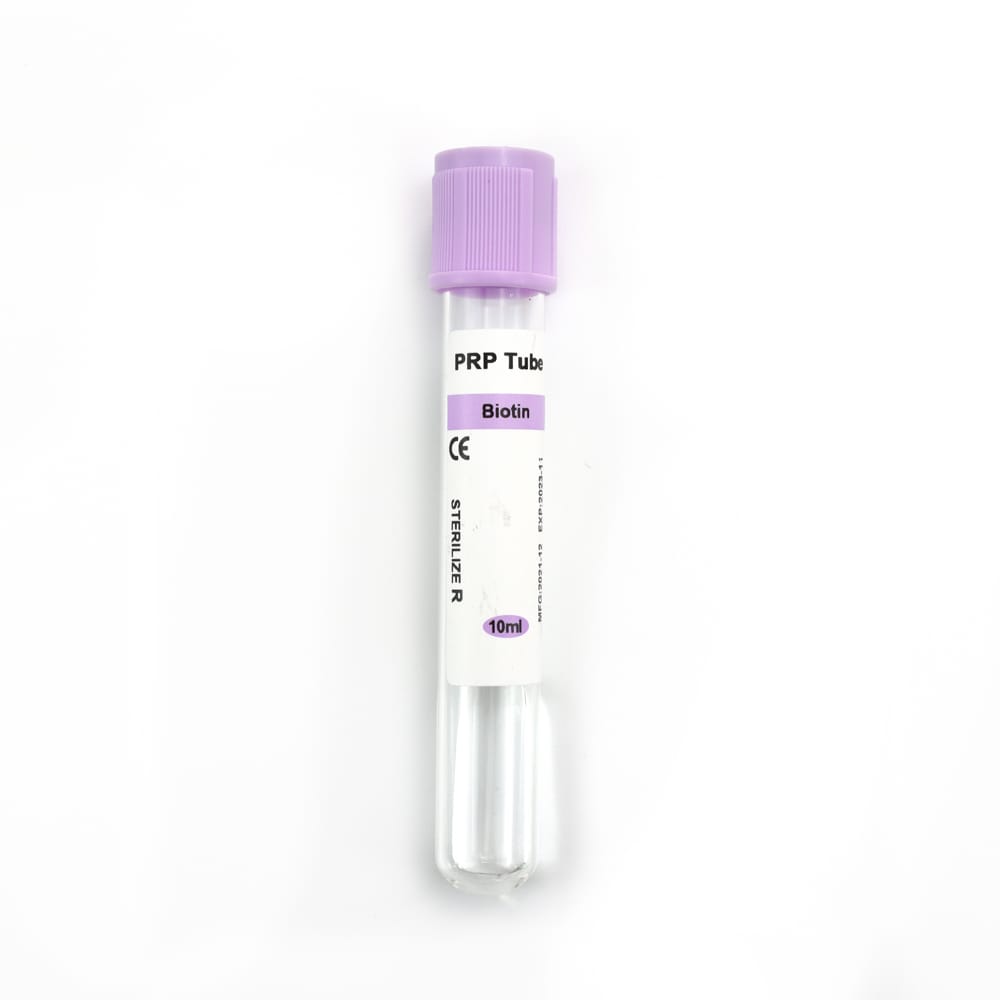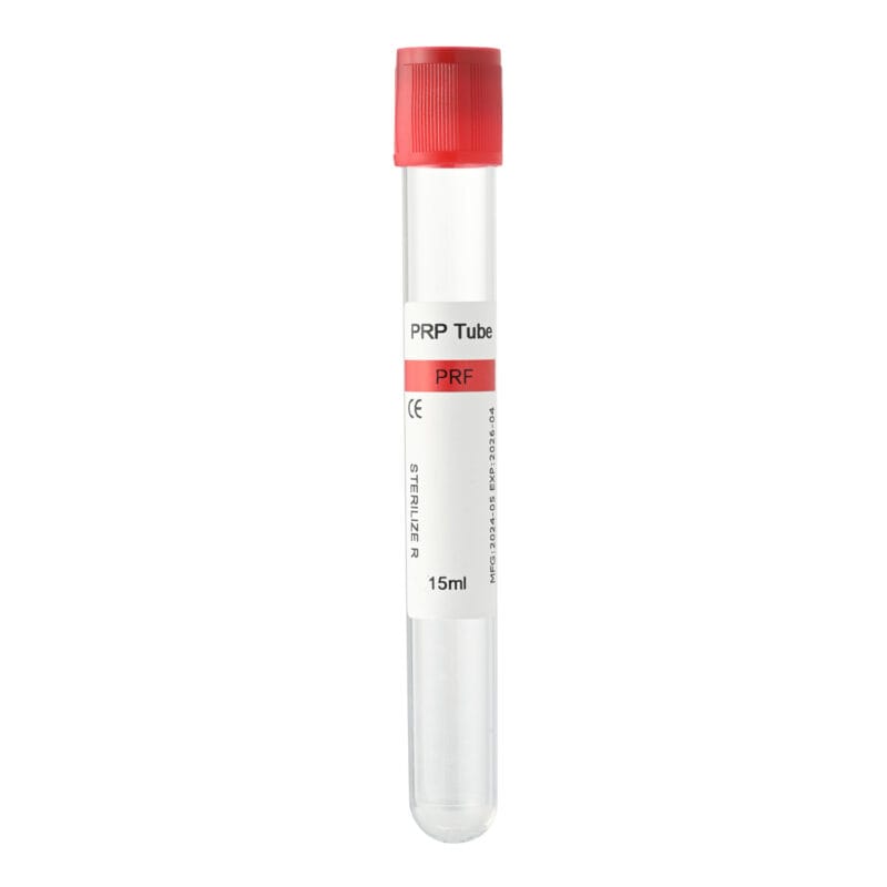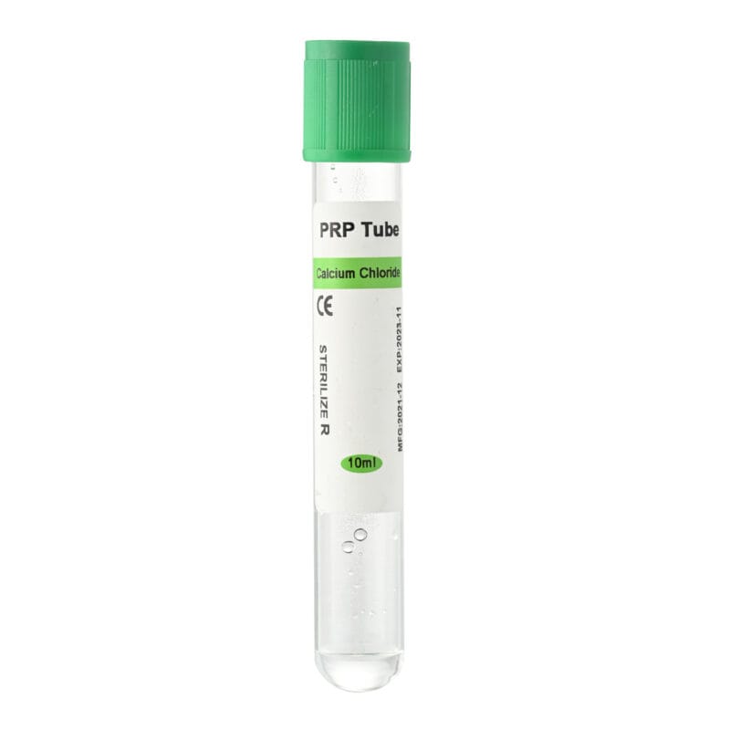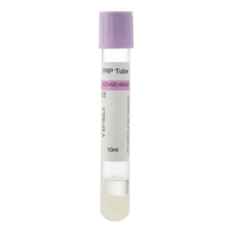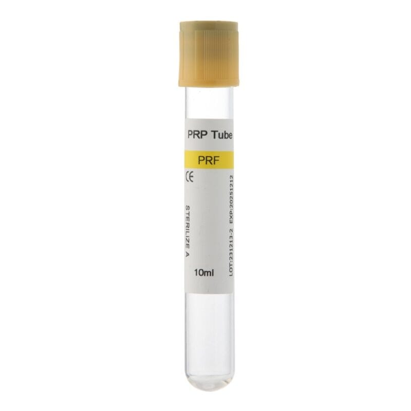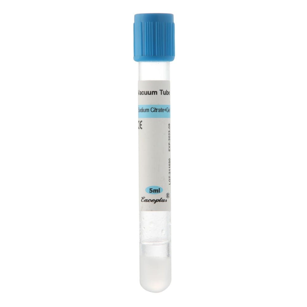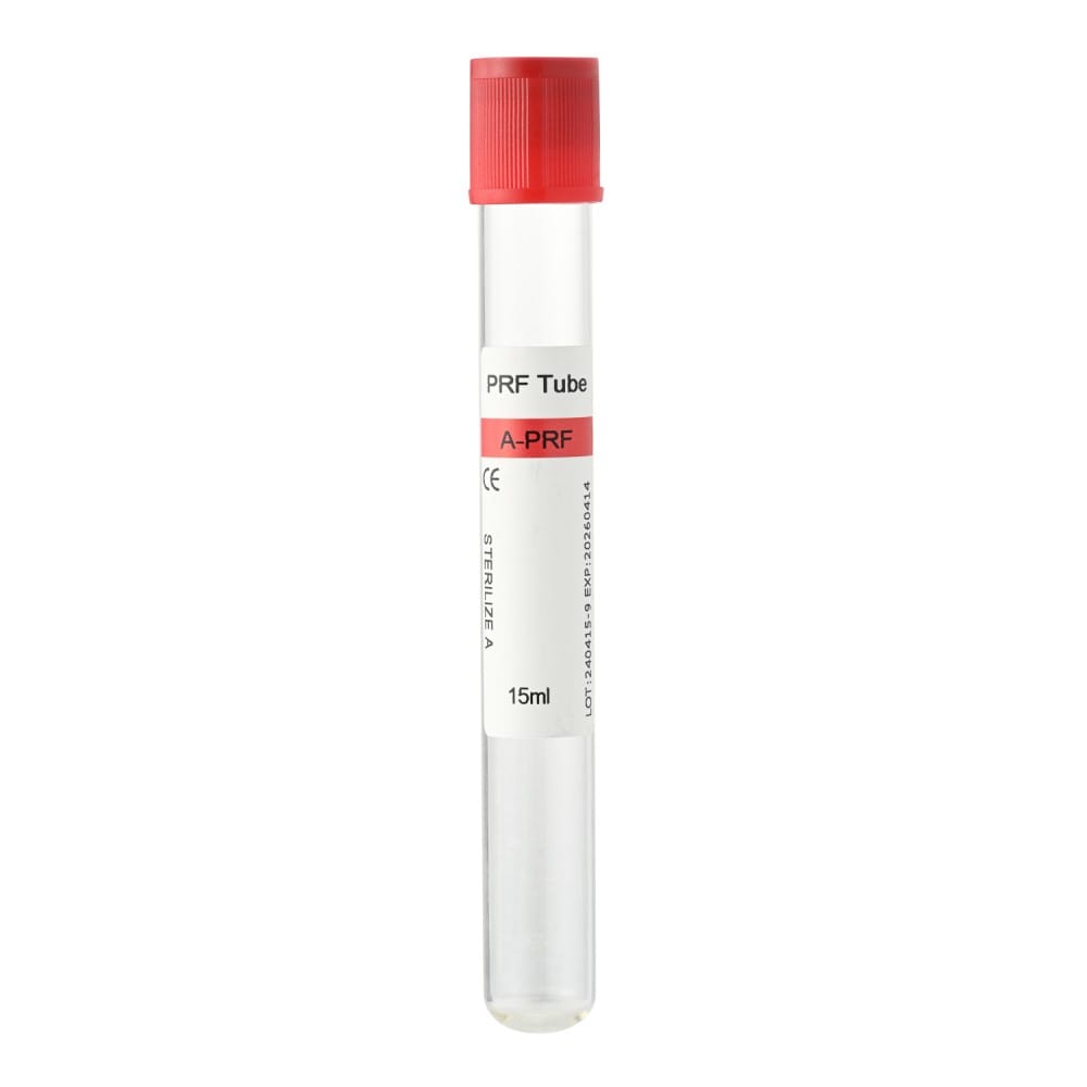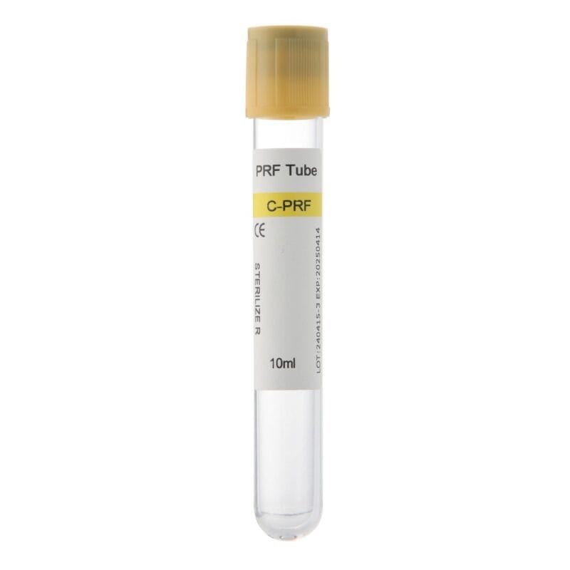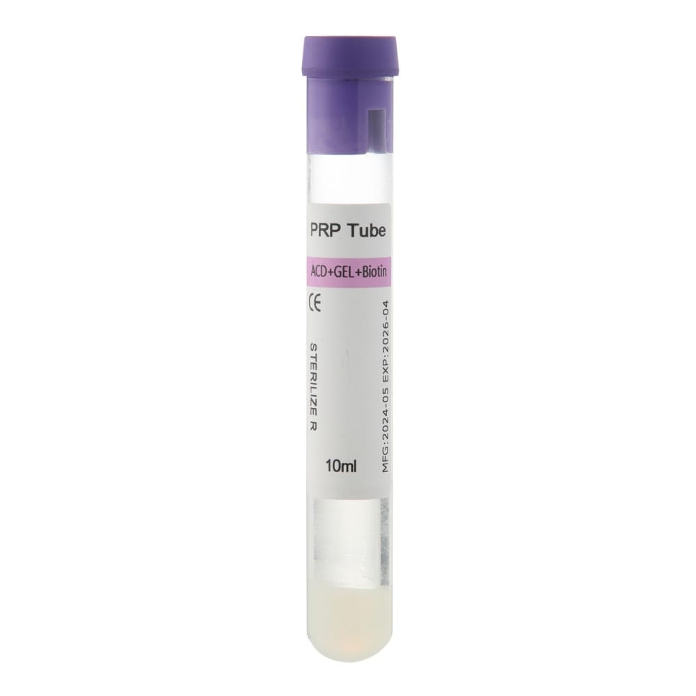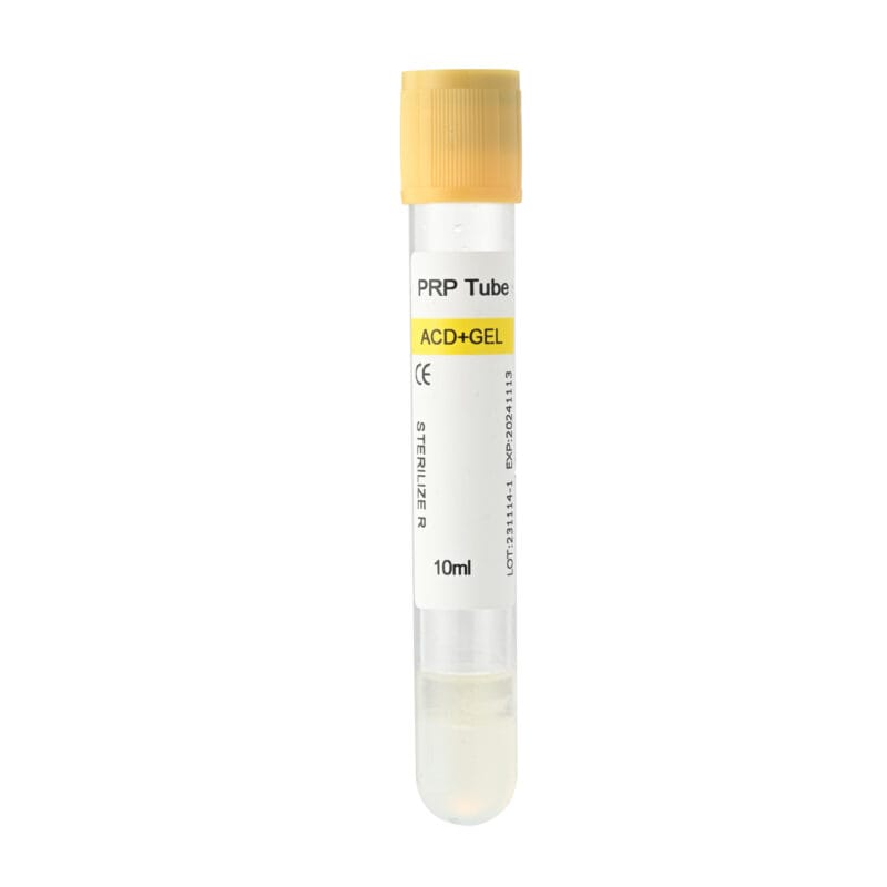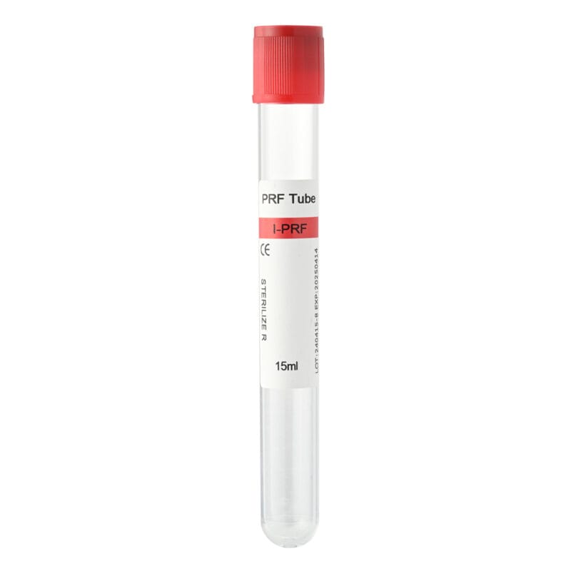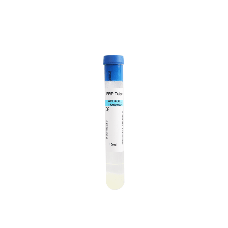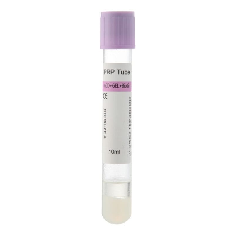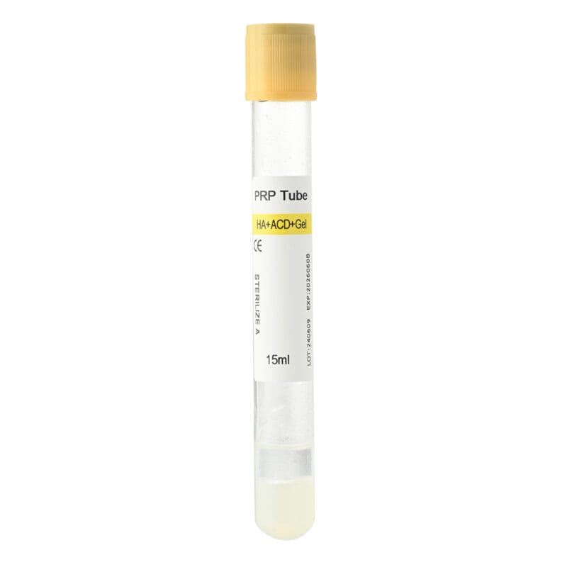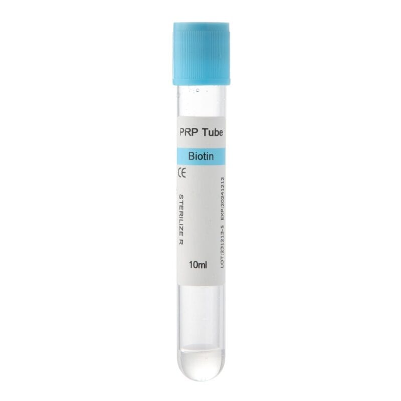
How to Make PRF Membranes is an important topic for dental professionals who use platelet-rich fibrin in regenerative procedures. These biological membranes come from a patient’s own blood and contain a three-dimensional fibrin network that holds platelets, leukocytes, and signaling molecules involved in tissue healing.
PRF membranes are commonly used as adjuncts in oral surgery, implant dentistry, and periodontal procedures. However, producing a consistent membrane involves more than collecting blood and running a centrifuge. The collection technique, tube characteristics, centrifugal force, and compression method can all influence the final material.
For dental clinics, understanding the preparation process helps improve workflow consistency. For medical distributors and purchasing teams, it also highlights why choosing appropriate PRF tubes matters. This guide explains how fibrin membranes form, outlines the main preparation steps, identifies common processing problems, and discusses practical considerations when selecting PRF collection tubes.
What Is a PRF Membrane and How Does It Work?
Platelet-rich fibrin (PRF) is an autologous blood-derived material containing a fibrin matrix, platelets, leukocytes, and various biological signaling molecules. Unlike conventional platelet-rich plasma preparation, many solid PRF protocols do not require anticoagulants or externally added clotting agents. Instead, blood coagulates during processing, creating a fibrin network that can be shaped into a membrane.
How Does a PRF Membrane Form?
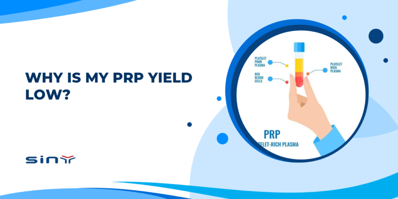
After blood enters a suitable collection tube, coagulation begins. Centrifugation separates blood components while the fibrin network develops. In a typical solid PRF preparation, the processed sample contains three recognizable regions:
- Upper region: A relatively clear, serum-rich fraction.
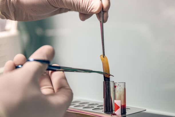
- Middle region: A yellowish fibrin clot containing platelets and leukocytes.
- Lower region: A concentrated red blood cell fraction.
The exact appearance and distribution of these regions depend on the preparation protocol, centrifugation conditions, and patient-related factors. Clinicians carefully separate the fibrin clot and compress it into a thin, flexible membrane.
The resulting material acts as a temporary biological scaffold. Its fibrin network can support cell attachment and organization, while incorporated platelets and leukocytes contribute signaling molecules associated with wound healing.
Why Are PRF Membranes Used in Dentistry?
Dental professionals use fibrin membranes in selected procedures involving soft-tissue healing and regenerative treatment. For example, clinicians may apply them during tooth extraction, periodontal surgery, or implant-related procedures. Their flexible structure allows them to adapt to irregular surgical sites.
However, PRF membranes are not interchangeable with every conventional barrier membrane. Their mechanical strength, degradation behavior, and space-maintaining properties differ from those of materials designed specifically for guided bone regeneration. Therefore, clinicians must select the appropriate material according to the procedure and treatment objectives.
How to Make PRF Membranes: Step-by-Step Guide
Preparing a fibrin membrane requires a controlled workflow because each stage can affect the structure and handling characteristics of the final material. The following steps describe the general principles of conventional solid PRF preparation. They are intended for professional education rather than as a substitute for a validated clinical protocol.
Blood collection and membrane preparation must be performed by appropriately trained healthcare professionals using suitable equipment and materials.
Step 1: Prepare the Equipment and PRF Tubes
Before collecting blood, organize the necessary equipment. Preparing everything in advance helps reduce delays between collection and centrifugation.
Typical materials include:
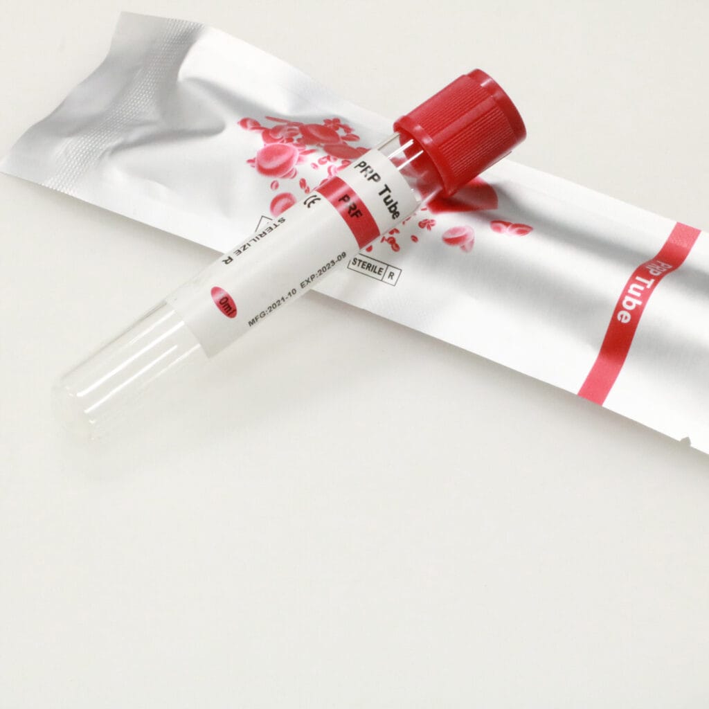
- PRF collection tubes suitable for the selected preparation protocol.
- A compatible medical centrifuge.
- Sterile blood collection equipment.
- Sterile forceps and handling instruments.
- A PRF compression box or membrane-forming device.
- Appropriate personal protective equipment.
Tube selection is especially important because the inner surface can influence contact activation and coagulation. Traditional solid PRF methods commonly use suitable glass collection tubes. Certain specially designed plastic tubes may also support fibrin formation, depending on their surface characteristics.
However, ordinary laboratory tubes should not automatically be considered suitable for clinical PRF preparation. Before use, verify the tube’s intended application, internal surface treatment, additives, sterility, and compatibility with the selected protocol.
Step 2: Collect Blood and Begin Processing Promptly
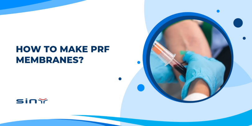
A qualified healthcare professional collects the required blood volume using an appropriate venipuncture technique. In conventional solid PRF protocols without anticoagulants, coagulation begins shortly after blood contacts the collection system. This makes timing an important part of the workflow.
If blood remains in the tube for too long before centrifugation, clot formation may progress prematurely. As a result, the fibrin matrix may develop differently from the intended protocol. Clinicians should therefore prepare the centrifuge before collection and follow the validated collection-to-centrifugation interval.
Tubes must also be positioned and balanced according to the centrifuge manufacturer’s instructions. When preparing several membranes, a consistent collection sequence can help minimize processing differences between samples.
Step 3: Centrifuge the Blood Using a Validated Protocol
Centrifugation separates blood components while the fibrin network develops. However, there is no single centrifugation setting suitable for every PRF system. Traditional leukocyte- and platelet-rich fibrin (L-PRF) protocols and advanced PRF techniques may use different centrifugal forces and processing times.
Two technical terms are particularly important:
- RPM (revolutions per minute): Describes how quickly the centrifuge rotor rotates.
- RCF (relative centrifugal force): Describes the centrifugal acceleration experienced by the sample, usually expressed as ×g.
RCF depends on both rotational speed and rotor radius. Therefore, two centrifuges operating at the same RPM may generate different centrifugal forces. Clinicians should not copy an RPM value from another device without confirming the corresponding RCF and protocol compatibility.
Rotor design, sample position, tube dimensions, and processing time may also influence the outcome. The safest approach is to use a validated protocol developed for the specific centrifuge, rotor, and collection tube combination.
Step 4: Separate the PRF Clot Carefully
After centrifugation, a fibrin-rich clot can usually be identified above the red blood cell fraction. The clot often appears yellowish and gelatinous. Using appropriate sterile instruments, clinicians carefully remove it from the tube. Gentle handling helps preserve the fibrin network and reduces unnecessary damage.
Particular attention is needed near the interface between the fibrin clot and red blood cell layer. Depending on the preparation protocol, platelets and leukocytes may be distributed unevenly throughout the clot, including near this interface. However, retaining more of the interface does not automatically produce a better membrane.
The objective is to follow the validated separation technique while limiting unwanted red blood cell contamination. Aggressive manipulation and unnecessary repeated handling should be avoided.
Step 5: Compress the PRF Clot Into a Membrane
Once separated, the fibrin clot can be converted into a thinner membrane using a dedicated PRF compression device. The clot is placed inside the sterile device, where controlled pressure removes excess fluid and produces a flatter structure.
Compression can influence membrane thickness, moisture retention, and handling characteristics. However, excessive or inconsistent pressure may change the physical properties of the material. For this reason, standardized compression is preferable to uncontrolled manual squeezing.
The appropriate compression time and pressure depend on the equipment and validated preparation method. After compression, the membrane should have sufficient structural integrity for its intended handling and clinical application. Its exact thickness, flexibility, and appearance may vary between protocols.
Step 6: Handle the Prepared Membrane Appropriately
Fresh PRF membranes require careful handling under appropriate aseptic conditions. Unnecessary exposure to air, contamination, excessive drying, and repeated manipulation should be avoided.
Some membrane-forming systems provide a protected environment for temporary handling. Nevertheless, this does not establish a universal storage duration. Clinicians should follow the validated handling instructions for their preparation system.
Whenever possible, the workflow should minimize unnecessary delays between preparation and clinical application. A consistent process helps reduce avoidable variation and supports reproducible membrane handling.
Key Factors That Affect PRF Membrane Quality
Understanding PRF membrane preparation is only the beginning. The final material can vary because of processing conditions, equipment characteristics, and biological differences between patients. Recognizing these factors helps clinicians identify problems without making arbitrary changes to their protocols.
Centrifugal Force and Processing Time
Centrifugal force influences blood component separation and the distribution of cells within the developing fibrin clot. Different protocols may produce fibrin matrices with different structural characteristics.
However, higher centrifugal force does not automatically mean better membrane quality. Similarly, lower-speed preparation is not universally superior. The appropriate settings depend on the intended PRF technique and the validated equipment configuration. For consistent results, clinics should use the same verified protocol rather than frequently changing centrifugation conditions.
Collection Timing and Coagulation
Blood collection timing is particularly important in protocols that do not use anticoagulants. Once coagulation begins, delays may influence the formation and organization of the fibrin network. Inconsistent timing can also make it difficult to compare results between preparation sessions.
A practical way to reduce variation is to establish a clear workflow before collection begins. This includes preparing the centrifuge, confirming tube compatibility, and ensuring trained staff are ready to process the samples.
Tube Surface and Material Characteristics
The collection tube can influence the early stages of coagulation. Glass surfaces can promote contact activation of the coagulation pathway, while plastic surfaces behave differently and may require specific treatments for certain solid PRF applications. Therefore, two tubes with the same nominal volume may not produce equivalent results.
Internal coatings and additives are also important. For example, a tube designed for anticoagulated blood collection should not be assumed suitable for a conventional solid PRF protocol that relies on natural coagulation. Product suitability must be verified rather than inferred from appearance.
Compression and Membrane Handling
Compression affects the physical form of the fibrin clot. A standardized compression device can help produce more consistent membrane dimensions and reduce differences caused by manual handling.
However, compression also removes fluid and may influence the distribution or retention of biological components. There is no universal rule that lighter or stronger compression always produces a better membrane. Instead, clinicians should follow an established method and avoid unnecessary changes.
Common Problems During PRF Membrane Preparation
| Problem | Possible cause | Practical consideration |
|---|---|---|
| Small or irregular clot | Collection delays or unsuitable processing conditions | Review timing and validated protocol |
| Weak membrane structure | Inconsistent fibrin formation or handling | Check tube suitability and processing workflow |
| Excessive red blood cell contamination | Improper clot separation | Review the separation technique |
| Overly dry membrane | Excessive compression or prolonged exposure | Standardize compression and handling |
| Different results between sessions | Variations in equipment, tubes, or timing | Maintain consistent preparation conditions |
| Unexpected clot appearance | Tube characteristics or patient-related variation | Review the full workflow before adjusting settings |
Visible differences do not always indicate a defective preparation. Patient-specific factors, including blood composition and physiological variation, may influence fibrin formation. Nevertheless, repeated problems should lead to a systematic review of the preparation process.
How to Choose the Right PRF Tubes for Membrane Preparation
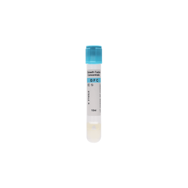
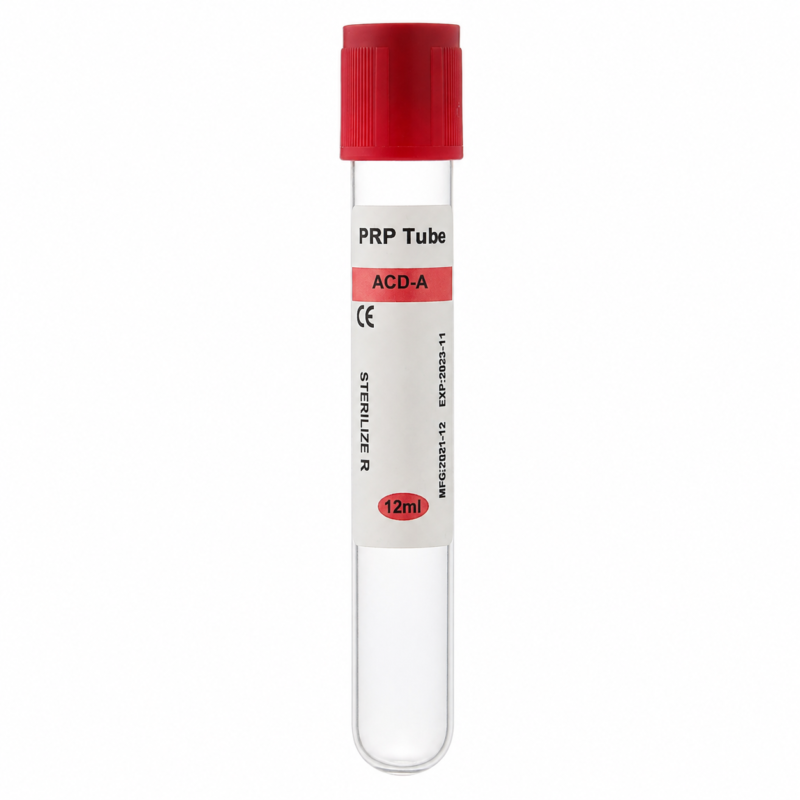
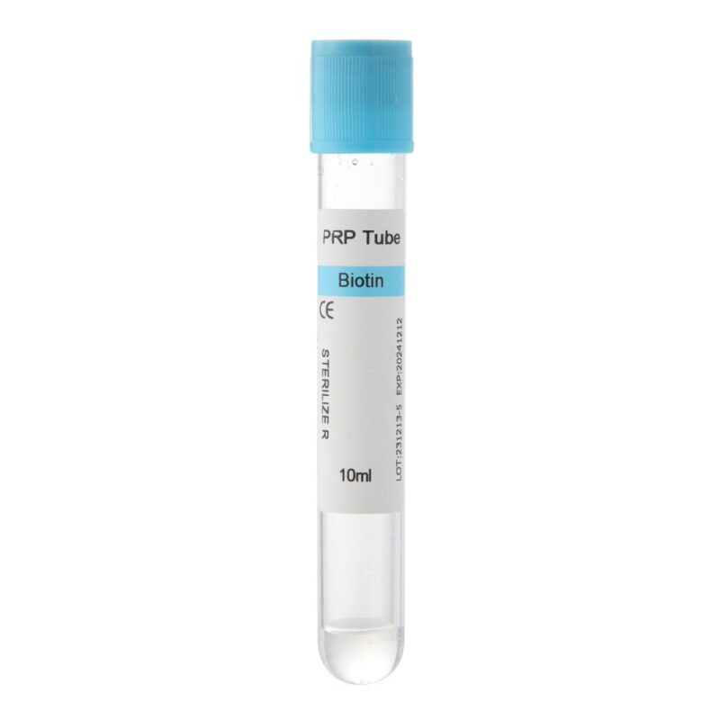
PRF tubes are essential consumables in the preparation process. Their characteristics can influence coagulation, clot formation, and workflow consistency. For dental clinics, choosing suitable tubes helps support a standardized preparation method. For distributors, understanding the differences between tube types is important when selecting products for professional customers.
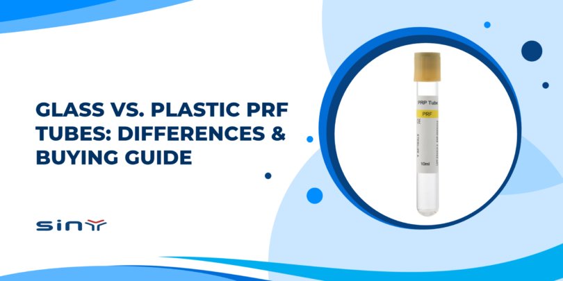
Glass PRF Tubes
Glass collection tubes have traditionally been used in several solid PRF protocols. Their surfaces can promote contact activation, supporting the coagulation process without anticoagulants.
However, glass tubes require appropriate handling because they may break if dropped or mishandled. Not every glass tube is suitable for clinical PRF preparation. The intended use, tube composition, sterility, and centrifuge compatibility must still be confirmed.
Plastic PRF Tubes
Plastic tubes are commonly used in medical laboratories because of their handling characteristics. However, plastic surfaces do not necessarily support coagulation in the same way as glass.
Some plastic PRF tubes are specifically designed with internal surface characteristics that support fibrin formation. Others may be intended for liquid fibrin preparation or different blood-processing applications. Therefore, buyers should verify the product’s intended use rather than assuming that all plastic PRF tubes are interchangeable.
Solid PRF Tubes vs. i-PRF Tubes
One important purchasing consideration is the distinction between conventional solid PRF and injectable PRF (i-PRF). Solid PRF preparation is intended to produce a fibrin clot that can be compressed into a membrane. In contrast, i-PRF protocols aim to obtain a liquid fibrin formulation that remains injectable for a limited period before coagulation progresses.
These applications involve different processing objectives. Although both belong to the broader PRF category, their tubes and preparation protocols should not automatically be treated as interchangeable. For clinics purchasing both product types, clear identification can help reduce selection errors.
PRF Tube Purchasing Comparison
| Purchasing factor | Solid PRF membrane preparation | Injectable PRF preparation |
|---|---|---|
| Main objective | Produce a fibrin clot for membrane formation | Obtain a liquid fibrin formulation |
| Expected material | Cohesive fibrin clot | Initially liquid preparation |
| Tube selection | Compatible with validated solid PRF protocol | Compatible with validated i-PRF protocol |
| Coagulation requirements | Support intended clot formation | Support the intended liquid-phase processing window |
| Processing conditions | Protocol-specific | Protocol-specific |
| Main purchasing concern | Clot formation and membrane preparation suitability | Liquid preparation suitability and handling time |
This comparison describes general preparation objectives rather than specifications for individual Siny PRP products.
What Should Dental Clinics and Distributors Check?
Before purchasing PRF collection tubes, buyers should evaluate several practical factors:
- Intended application: Confirm whether the product is designed for solid PRF, i-PRF, or another preparation method.
- Tube material and surface characteristics: Understand how the collection surface interacts with the intended coagulation process.
- Additives: Identify any anticoagulants, clot activators, separation gels, or other substances. Their presence may affect suitability for a particular protocol.
- Centrifuge compatibility: Verify tube dimensions, rotor compatibility, and supported processing conditions.
- Sterility and packaging: Review the relevant product information and handling requirements.
- Product consistency: Evaluate whether the supplier can provide stable product characteristics across repeat orders.
For distributors, accurate product descriptions are especially important. A tube should not be marketed for a particular clinical preparation simply because it resembles another product. Instead, product selection should be based on documented intended use and compatibility with the customer’s workflow.
Clinical Applications and Practical Considerations
PRF membranes are used or investigated in several dental procedures. Their biological characteristics make them useful as adjunctive materials, although clinical outcomes depend on the indication and treatment approach.
Tooth Extraction and Socket Management
After tooth extraction, clinicians may place a fibrin clot or membrane in selected surgical sites. The fibrin network provides a temporary biological matrix that may support early healing processes.
However, PRF does not guarantee improved outcomes in every extraction case. Its clinical value depends on the treatment protocol, patient characteristics, and surgical conditions.
Dental Implant Procedures
In implant dentistry, fibrin membranes may be used as adjuncts during selected soft-tissue or bone-related procedures. Their flexible structure allows them to adapt to irregular surfaces.
However, PRF membranes generally do not provide the same mechanical barrier or space-maintaining function as conventional guided bone regeneration membranes. They should therefore be selected according to the specific surgical objective.
Periodontal Procedures
PRF has also been investigated as an adjunct in periodontal regeneration and soft-tissue healing. Its fibrin matrix and associated biological components may contribute to the local healing environment.
Nevertheless, clinical results vary across indications, and the available evidence should guide treatment decisions.
Building a Consistent Clinical Workflow
For clinics that regularly prepare PRF membranes, consistency is a practical priority. A standardized workflow should address blood collection, centrifugation, clot separation, compression, and handling.
Staff training is equally important because small differences in timing or technique may influence preparation. Reliable consumable selection also supports workflow consistency. By using appropriately selected tubes and following validated procedures, clinics can reduce avoidable variation and make routine membrane preparation more predictable.
Frequently Asked Questions About PRF Membranes
1. What is a PRF membrane made of?
A PRF membrane is produced from a patient’s own blood. It contains a fibrin network with platelets, leukocytes, and associated biological components. Its exact composition depends on the preparation protocol.
2. How long does PRF membrane preparation take?
Preparation time depends on blood collection, centrifugation, clot separation, and compression. Different systems use different protocols, so there is no universal preparation time.
3. Do PRF membranes require anticoagulants?
Conventional solid PRF protocols generally use blood collected without anticoagulants. Natural coagulation allows the fibrin network to develop. Other platelet-concentrate techniques may have different requirements.
4. Can plastic tubes be used to prepare PRF membranes?
Certain plastic tubes are designed for solid PRF preparation. However, suitability depends on their internal surface characteristics, additives, intended use, and compatibility with the selected protocol.
5. Why is my PRF membrane thin or weak?
Possible causes include processing delays, unsuitable tube characteristics, inconsistent centrifugation, or excessive compression. Patient-related biological variation may also influence the result.
6. What is the difference between a PRF clot and a PRF membrane?
A PRF clot is the fibrin-rich material obtained after processing blood. A PRF membrane is formed by compressing that clot into a thinner structure suitable for selected clinical applications.
7. Can i-PRF be used to make a conventional PRF membrane?
Injectable PRF is prepared to obtain an initially liquid formulation. Although it subsequently coagulates, it is not automatically equivalent to a conventional solid PRF clot intended for compression into a membrane.
8. Are PRF membranes suitable for every dental procedure?
No. Their suitability depends on the clinical indication, treatment objectives, and available evidence. PRF membranes may serve as biological adjuncts, but they do not replace every conventional regenerative material.
Conclusion
Learning how to make PRF membranes involves understanding the entire preparation process. Blood collection, centrifugation, clot separation, and compression all contribute to the characteristics of the final fibrin material.
For dental professionals, consistent preparation methods help reduce avoidable variation. For medical buyers, selecting appropriate collection tubes is equally important. Tube material, internal surface characteristics, intended application, and equipment compatibility should all be considered before purchasing.
Siny PRP supplies glass and plastic PRF tubes, including conventional PRF and i-PRF product options, for professional medical and dental markets. Explore our products at sinyprp.com or contact our team to discuss PRF tube options and purchasing requirements for your clinic or distribution business.