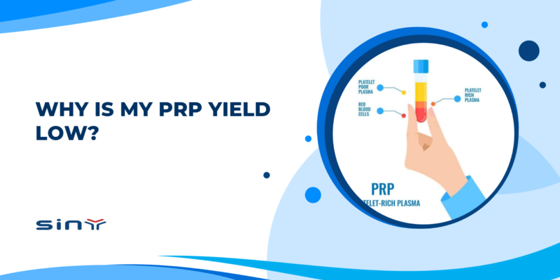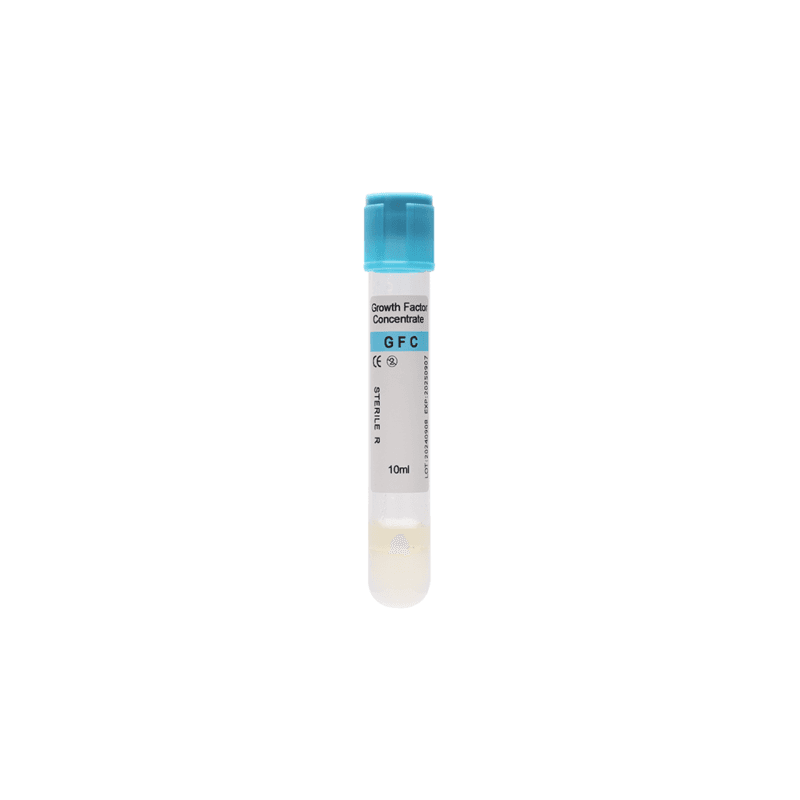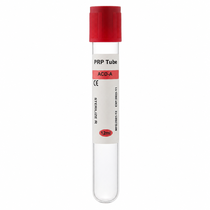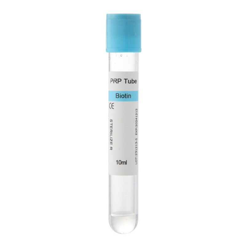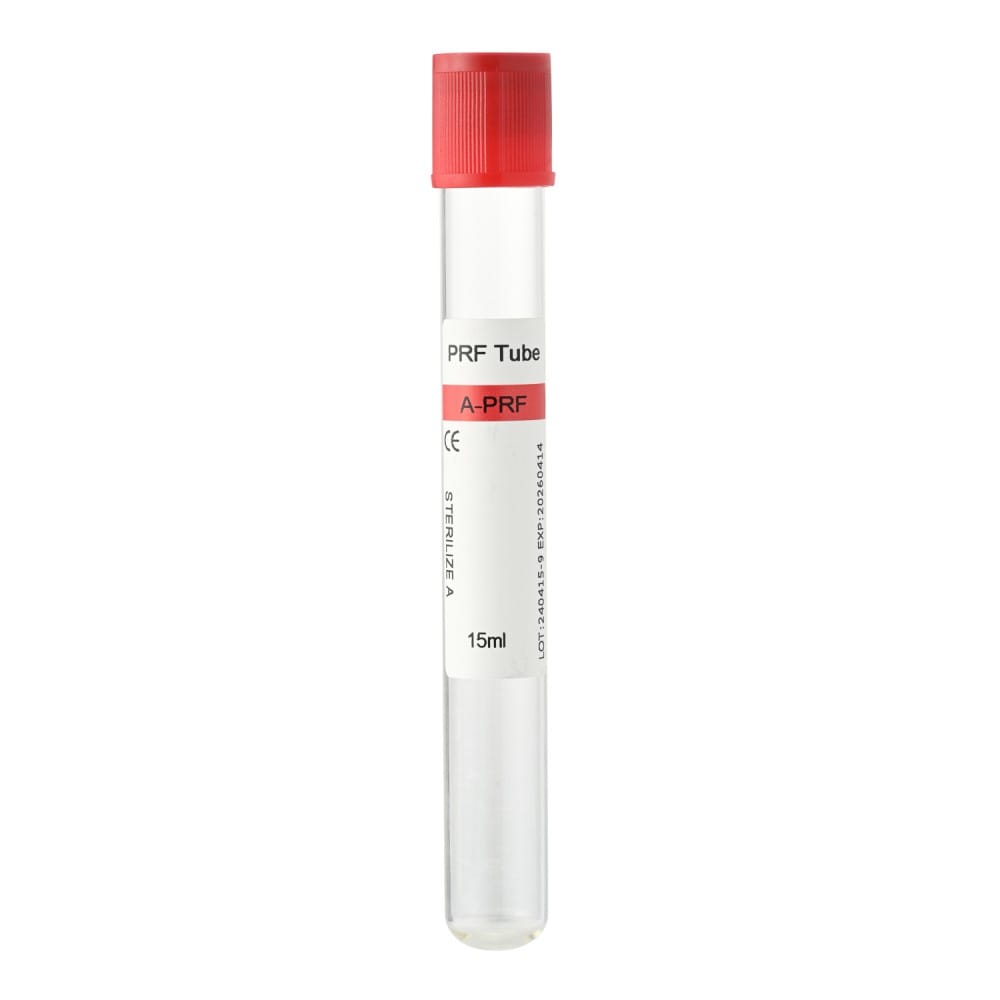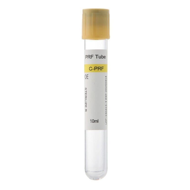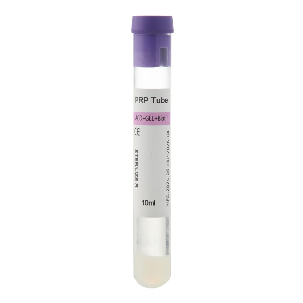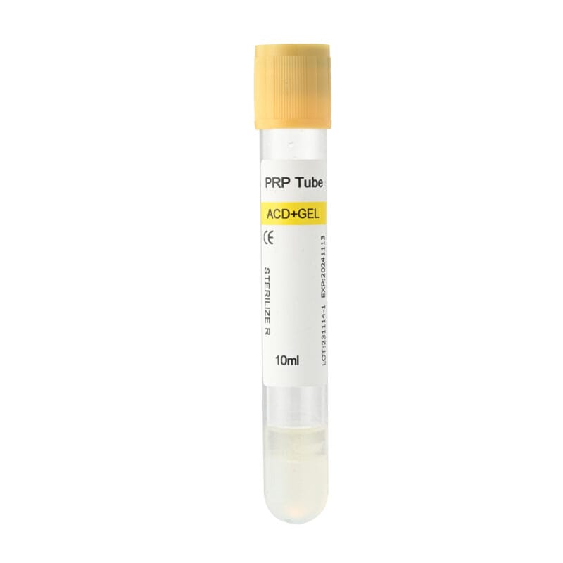
Why Is My PRP Yield Low? This is a common question among clinics and medical professionals who prepare platelet-rich plasma (PRP) for aesthetic, orthopedic, dental, and other clinical applications.
Sometimes, a blood sample produces less PRP than expected. In other cases, the collected plasma volume appears normal, but the platelet concentration is lower than anticipated. These situations may look similar, yet their causes can be quite different.
Low PRP output can result from incorrect centrifugation settings, insufficient blood collection, anticoagulant problems, platelet loss during extraction, or an unsuitable PRP tube configuration. Patient-related blood characteristics can also influence the final result.
Understanding these factors helps clinics identify preparation problems, reduce unnecessary platelet loss, and maintain a more consistent workflow.
This guide explains what low platelet yield means, the most common causes, practical troubleshooting methods, and how PRP collection tube selection can affect preparation efficiency.
What Does Low PRP Yield Actually Mean?
Before adjusting centrifugation settings or changing collection tubes, it is important to identify what is actually low: PRP volume, platelet concentration, or platelet recovery.
These measurements describe different aspects of PRP preparation.
PRP Volume vs. Platelet Concentration
PRP volume refers to the amount of platelet-rich plasma collected after processing, usually measured in milliliters (mL).
Platelet concentration describes the number of platelets present in a given volume of plasma, typically expressed as platelets per microliter (µL).
A larger PRP volume does not necessarily contain more platelets per microliter. Similarly, a smaller preparation may have a higher platelet concentration.
Consider two hypothetical PRP samples prepared from the same initial blood volume and baseline platelet count.
| Measurement | Sample A | Sample B |
|---|---|---|
| Initial whole blood volume | 10 mL | 10 mL |
| Baseline platelet count | 200,000/µL | 200,000/µL |
| Final PRP volume | 2 mL | 4 mL |
| Final platelet concentration | 800,000/µL | 400,000/µL |
| Platelet concentration factor | 4× | 2× |
| Calculated platelet recovery | 80% | 80% |
Both samples contain approximately the same total number of recovered platelets, despite their different volumes and concentrations.
This example illustrates why evaluating PRP preparation by volume alone can be misleading.
What Is Platelet Recovery Rate?
Platelet recovery measures the proportion of platelets from the original blood sample that are retained in the final PRP preparation.
It can be estimated using the following formula:
Platelet Recovery (%) = (Final PRP Platelet Count × Final PRP Volume) ÷ (Baseline Whole Blood Platelet Count × Initial Whole Blood Volume) × 100
Platelet counts and volumes must use consistent units. The calculation should also account for anticoagulant-related dilution and any relevant sample-processing adjustments.
A high platelet concentration does not automatically indicate high platelet recovery. Some preparation methods concentrate platelets into a smaller plasma volume while losing a portion of the original platelet population.
The desired concentration and recovery also depend on the intended PRP formulation. More concentrated PRP is not necessarily more effective for every clinical application.
What Causes Low PRP Yield?
Poor platelet recovery can originate before, during, or after centrifugation. Identifying the stage where platelets are lost is usually more useful than immediately changing the centrifuge speed.
Incorrect PRP Centrifugation Speed or Time
Centrifugation separates blood components according to their physical properties.
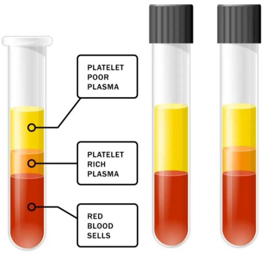
During processing, red blood cells generally move toward the bottom of the tube, while plasma remains above the denser cellular fraction. Platelet distribution depends on centrifugal force, processing duration, and the preparation method.
If the applied force or processing time is insufficient, blood components may not separate as intended.
Conversely, excessive centrifugal force can move platelets into fractions that are not collected by a particular protocol. Under some conditions, unsuitable processing may also affect platelet integrity.
An important distinction is the difference between RPM and RCF.
- RPM (Revolutions Per Minute): Measures rotor rotation speed.
- RCF (Relative Centrifugal Force): Measures the centrifugal acceleration applied to the sample, expressed as multiples of gravitational acceleration (×g).
Two centrifuges operating at identical RPM settings can generate different RCF values because their rotor radii differ.
Therefore, copying RPM settings between centrifuges may lead to inconsistent PRP separation.
The appropriate centrifugal force depends on the rotor, collection tube, processing method, and intended platelet-containing fraction.
Insufficient Blood Collection Volume
The initial blood volume determines how many platelets are available for processing.
For example, collecting less blood generally reduces the total number of available platelets when baseline platelet concentrations are comparable.
Underfilling a PRP tube can introduce another problem.
Some collection tubes contain a predetermined amount of anticoagulant. If the blood volume is lower than intended, the blood-to-anticoagulant ratio may change.
This can affect sample dilution and potentially influence the preparation process.
For reliable results, the collected blood volume should remain within the tube manufacturer’s specified filling range.
Improper Anticoagulant Mixing
Anticoagulants help prevent blood from clotting during PRP preparation.
Common formulations include acid citrate dextrose (ACD) and sodium citrate. Both work primarily by binding calcium, which is required for normal coagulation.
If blood is not adequately mixed with the anticoagulant, localized clotting or platelet aggregation may occur.
Platelets trapped in clots may not be recovered in the final PRP fraction.
However, vigorous shaking is not an appropriate solution. Excessive mechanical agitation may damage blood components or promote unwanted platelet activation.
Gentle mixing according to the tube instructions helps maintain a more consistent sample.
Poor Blood Component Separation
After centrifugation, the separation between plasma and cellular components should be appropriate for the selected preparation system.
If the layers are poorly defined, collecting the intended platelet-rich fraction becomes more difficult.
Possible causes include unsuitable centrifugation conditions, sample handling problems, or incompatibility between the tube and centrifuge.
For gel-based PRP tubes, separator position is particularly important.
The gel is designed to form a barrier between selected blood fractions. However, platelet distribution relative to that barrier varies with tube design and processing conditions.
A clearly visible separator does not, by itself, confirm that platelet recovery is optimal.
Platelet Loss During Plasma Extraction
Platelet loss can occur even when centrifugation has produced an appropriate separation.
In some preparation systems, platelets accumulate near the interface between plasma and the cellular fraction.
Collecting only the upper plasma may leave behind a substantial portion of the available platelets.
On the other hand, aspirating too close to the red blood cell layer may introduce unwanted cellular contamination.
The correct extraction location depends on the intended PRP composition and whether the system produces leukocyte-rich or leukocyte-poor PRP.
Careful aspiration and adherence to the system-specific collection protocol are essential.
How to Improve PRP Yield and Platelet Recovery
Improving PRP preparation requires a systematic approach. Changing several processing variables simultaneously can make it difficult to identify which adjustment actually improves the result.
The following steps help address common preparation problems.
Step 1: Standardize Blood Collection
Begin by confirming that the blood sample meets the collection requirements.
Use the intended tube filling volume, mix the blood with the anticoagulant as instructed, and avoid unnecessary delays before processing.
Check for visible clotting or other sample abnormalities before centrifugation.
Patient-related factors, including baseline platelet count and hematocrit, can also affect the final preparation.
If results vary significantly between patients despite consistent handling, baseline blood characteristics may explain part of the difference.
Step 2: Verify Centrifugation Parameters
Confirm that the centrifuge settings match the selected PRP preparation system.
Important variables include:
- Relative centrifugal force (RCF)
- Centrifugation duration
- Rotor radius and configuration
- Single-spin or double-spin protocol
- Tube compatibility and positioning
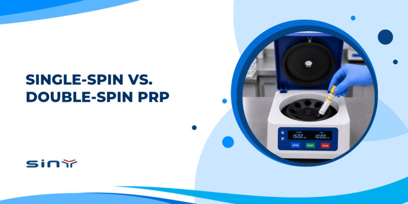
Single-spin methods use one centrifugation cycle to separate the desired blood fractions.
Double-spin methods introduce an additional processing step, often to concentrate platelets within a smaller plasma volume.
Although double-spin processing can increase platelet concentration under certain conditions, it does not automatically provide greater total platelet recovery.
Additional handling may also introduce opportunities for platelet loss.
For this reason, clinics should follow an established, validated preparation protocol rather than assuming that more centrifugation produces better PRP.
Step 3: Improve PRP Extraction Accuracy
Once centrifugation is complete, handle the tube carefully to avoid disturbing the separated layers.
Identify the platelet-containing fraction specified by the preparation system before beginning extraction.
Use controlled aspiration and avoid collecting unwanted blood components.
Some protocols also require gentle resuspension of the platelet-containing fraction before final collection or laboratory measurement.
This helps reduce uneven platelet distribution within the sample.
When platelet counts are available, measuring the final preparation provides a more reliable assessment than judging plasma color or volume alone.
Step 4: Identify the Problem Using a Troubleshooting Table
The following table summarizes common preparation problems and practical corrective actions.
| Observed Problem | Possible Cause | Recommended Action |
|---|---|---|
| Low collected plasma volume | Insufficient blood collection or high hematocrit | Verify the intended fill volume and review sample characteristics |
| Low platelet concentration | Inappropriate separation or extraction method | Review RCF, centrifugation time, and collection fraction |
| Visible blood clots | Inadequate anticoagulant mixing or delayed handling | Check collection and mixing procedures |
| Poorly defined separation layers | Unsuitable processing conditions | Confirm centrifuge and tube compatibility |
| Platelets remaining in discarded fractions | Incomplete recovery of the intended platelet-rich layer | Review the extraction protocol and assess platelet distribution |
| Red blood cell contamination | Disturbance of the cellular interface | Improve aspiration control |
| Inconsistent results between preparations | Variable handling or patient blood characteristics | Standardize procedures and compare preparation records |
Visual observations are useful for initial troubleshooting, but they cannot reliably establish platelet concentration or recovery.
Where practical, laboratory platelet counts should be used to evaluate preparation performance.
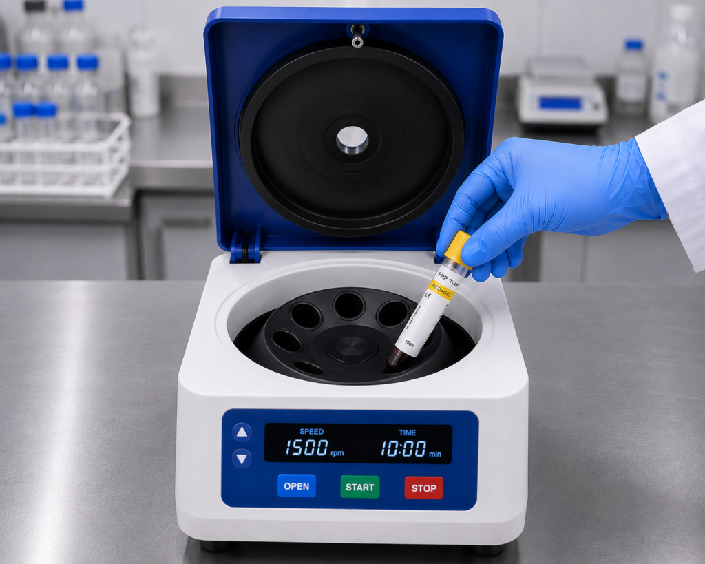
Can PRP Tube Selection Affect Platelet Yield?
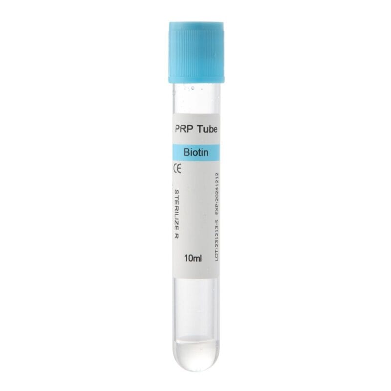
The PRP collection tube is an important part of the preparation system.
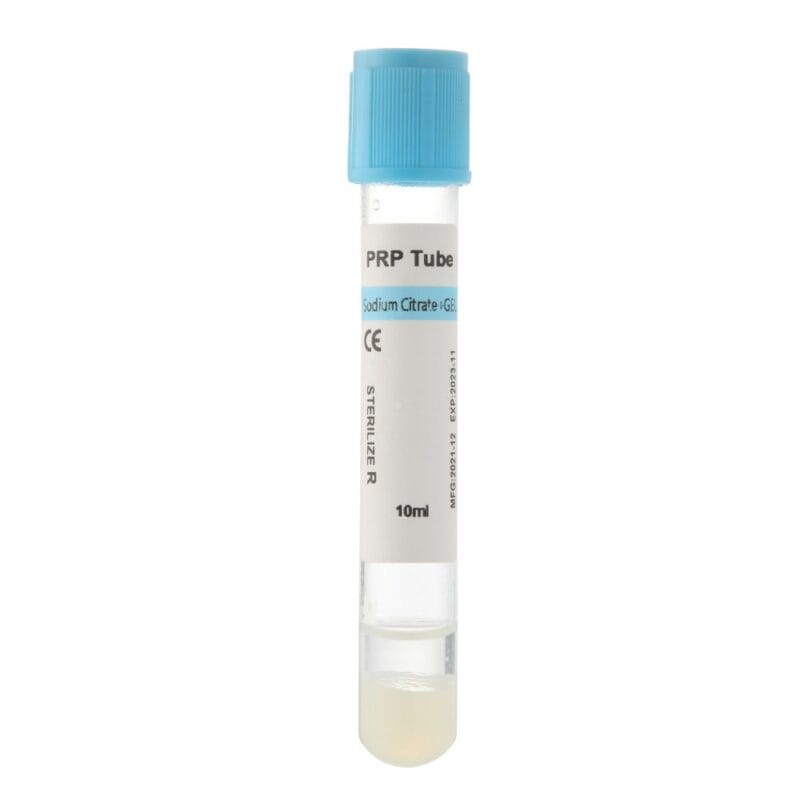
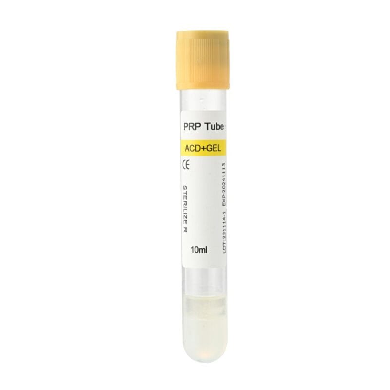
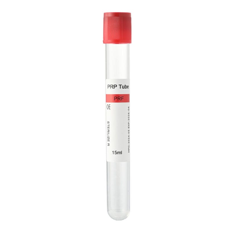
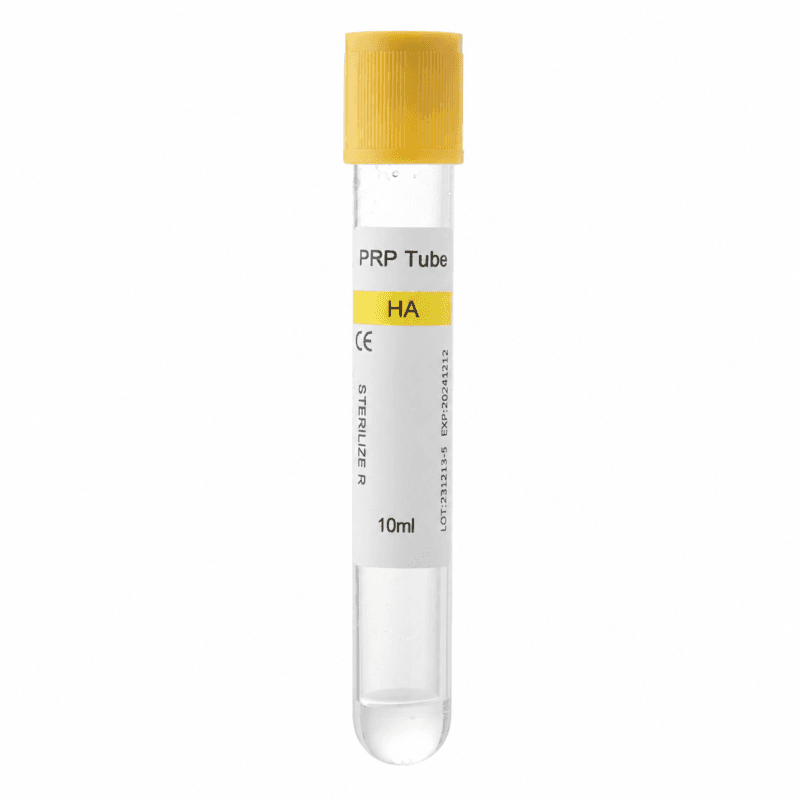
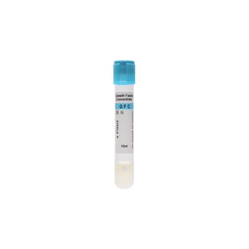
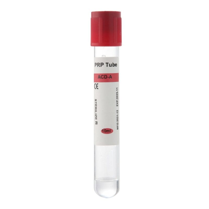
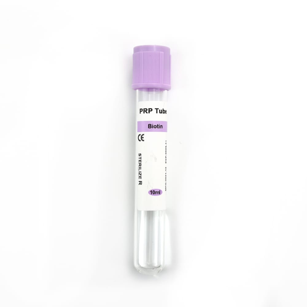
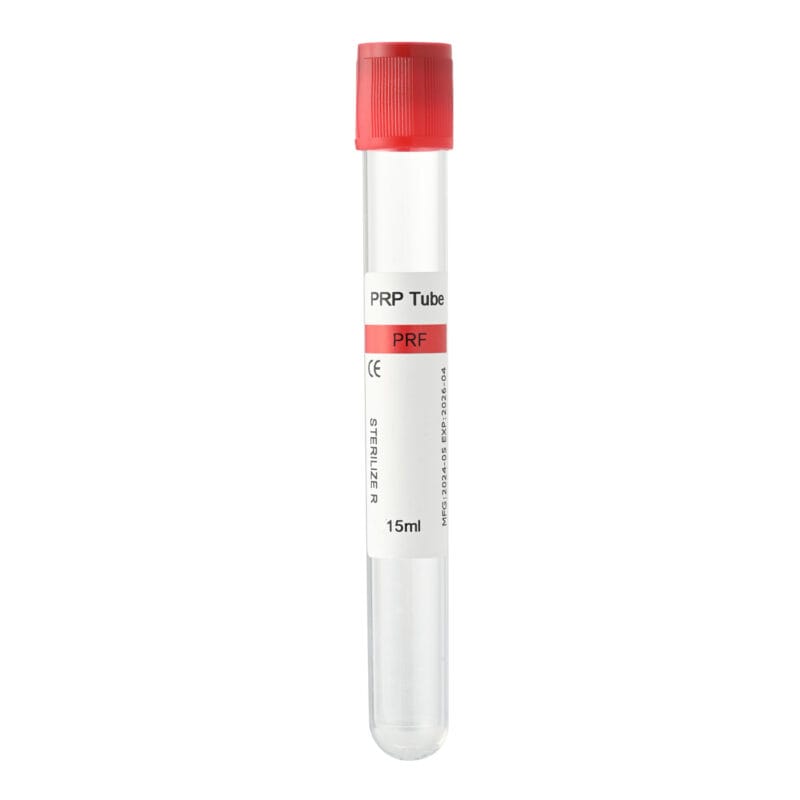
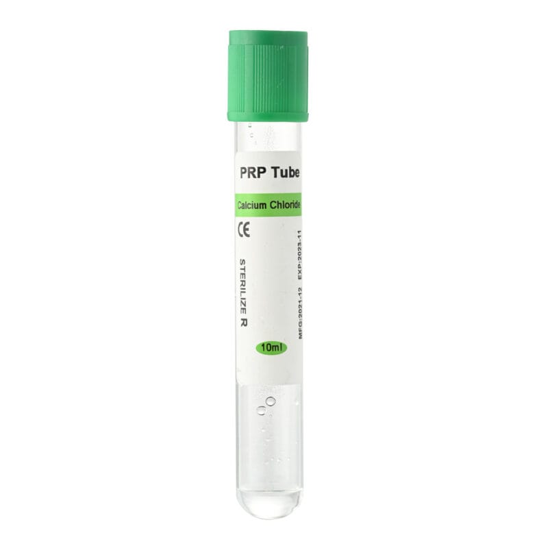
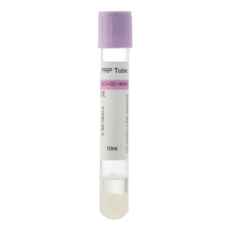
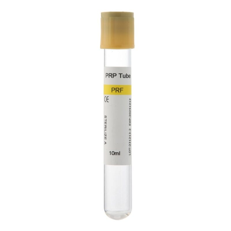
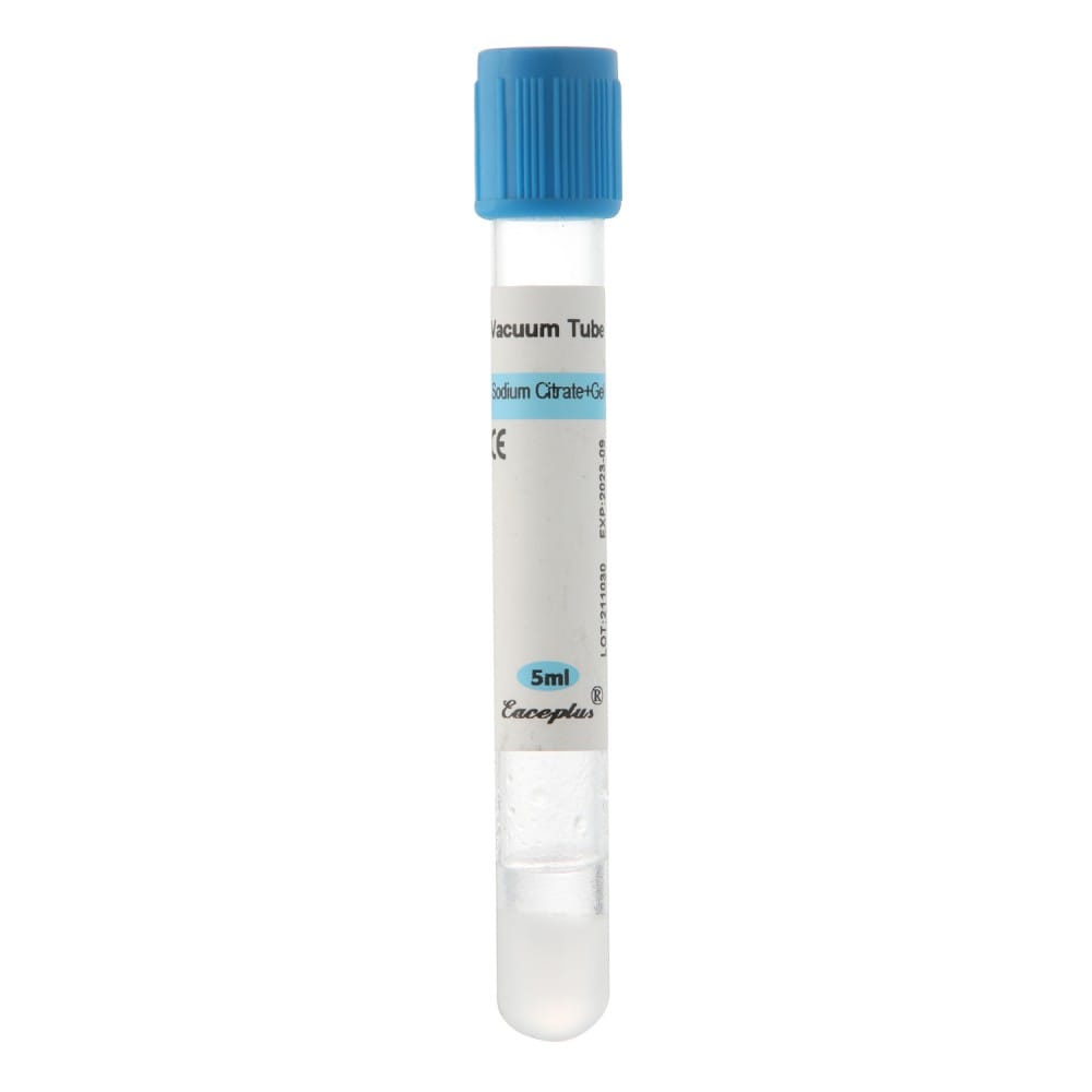
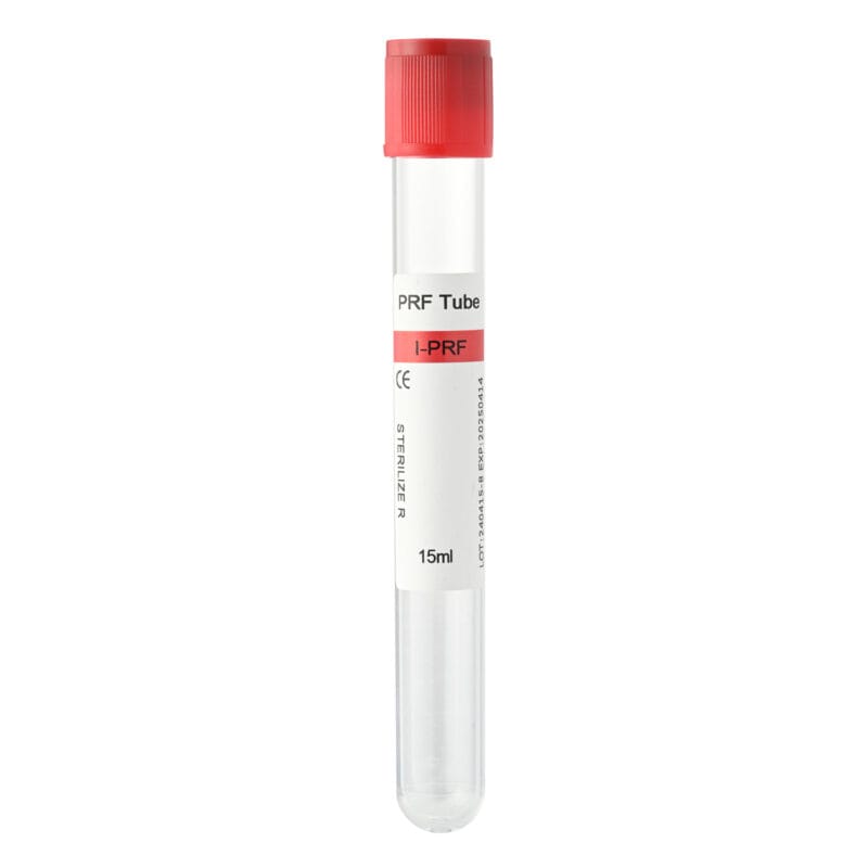
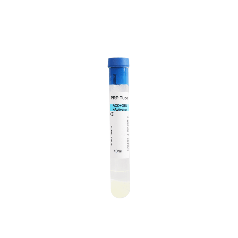
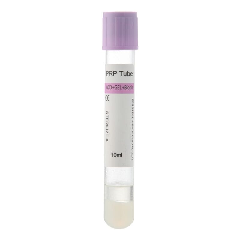
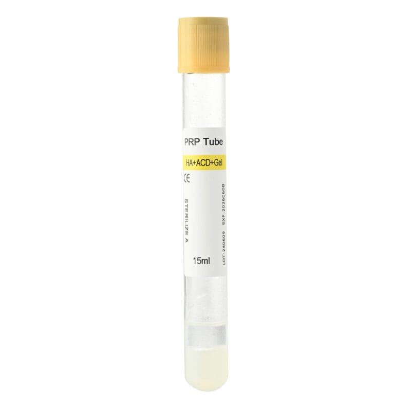
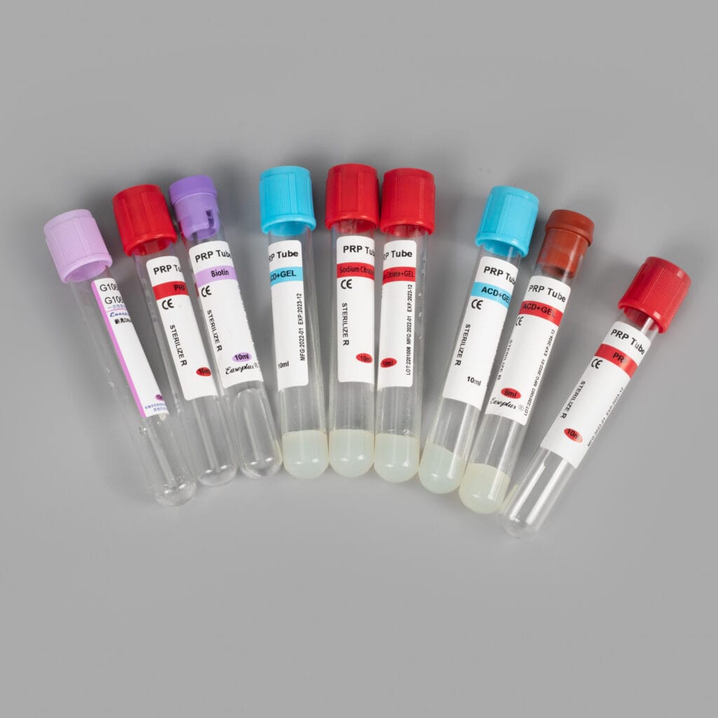
Tube design, anticoagulant formulation, separator configuration, and intended filling volume can influence how blood components separate and how the platelet-containing fraction is collected.
However, no tube configuration guarantees high platelet recovery under every processing condition.
Anticoagulant Selection
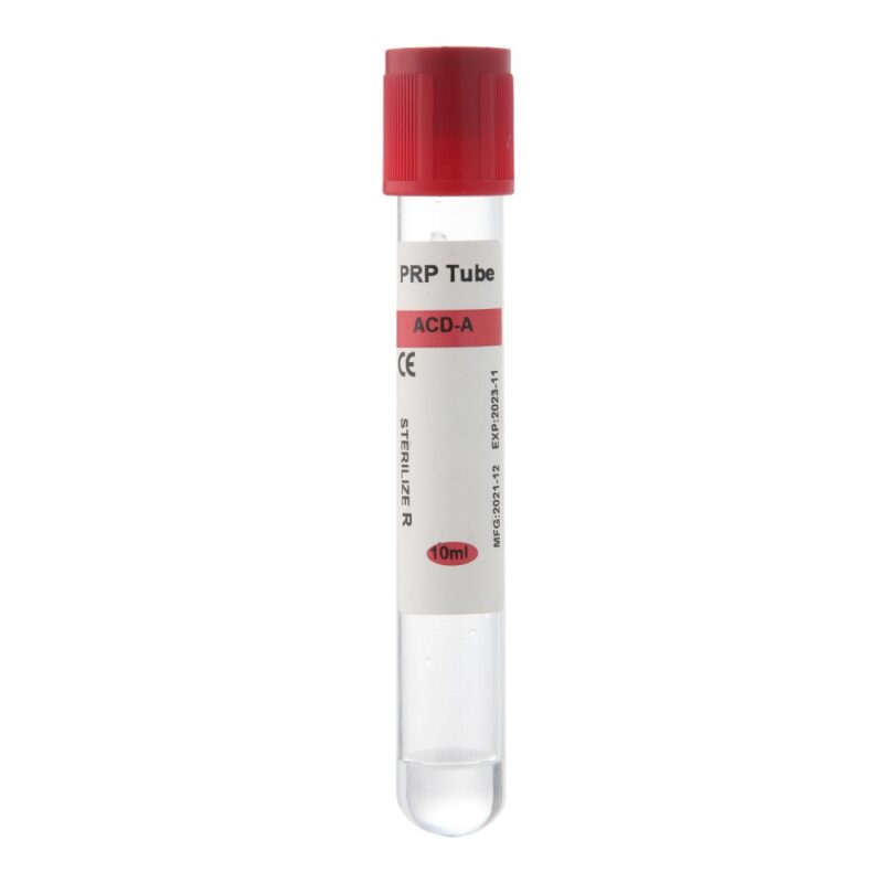
ACD and sodium citrate are commonly used in PRP preparation.
Both help prevent coagulation through calcium binding, but their formulations and dilution effects differ.
The appropriate anticoagulant depends on the preparation system and intended protocol.
For medical buyers, the main consideration is whether the additive formulation is compatible with the collection method.
Choosing a different anticoagulant does not automatically increase platelet concentration.
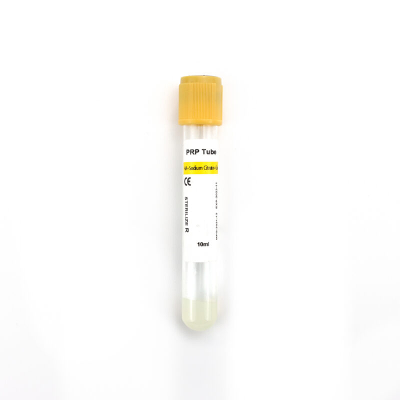
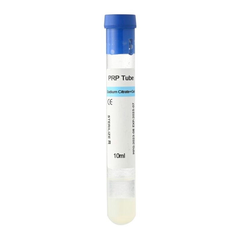
Gel vs. Non-Gel PRP Tubes
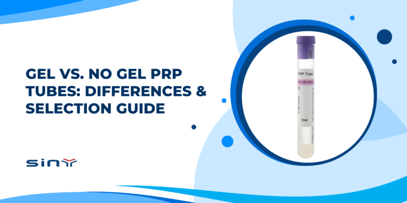
Gel-based and non-gel PRP tubes use different approaches to blood component separation.
Gel-based systems incorporate a physical separator, while non-gel systems rely on the separation and collection method without the same barrier.
Both configurations can be suitable for PRP preparation when used with an appropriate protocol.
| Feature | Gel PRP Tubes | Non-Gel PRP Tubes |
|---|---|---|
| Separation design | Uses a gel separator | No gel barrier |
| Plasma collection | Depends on separator position and tube design | Depends on the intended extraction method |
| Platelet recovery | System-dependent | System-dependent |
| Handling | May simplify separation in compatible systems | May allow flexible collection of selected fractions |
| Main consideration | Separator and protocol compatibility | Extraction accuracy and processing method |
A gel separator may simplify handling, but its presence does not guarantee superior platelet recovery.
Similarly, non-gel tubes do not automatically produce higher platelet concentrations.
The appropriate choice depends on the intended PRP composition, centrifugation protocol, and operator workflow.
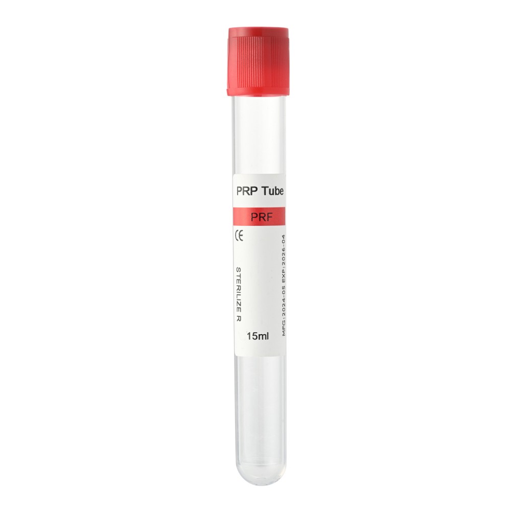
PRP Tube Capacity and Filling Volume
Tube capacity should match the intended blood collection volume.
A larger collection tube may accommodate more blood, but it does not necessarily improve the percentage of platelets recovered.
For tubes containing anticoagulant, correct filling is particularly important because the additive quantity is generally designed for a specified blood volume.
Clinics should therefore select tube capacities based on their preparation requirements rather than assuming that larger tubes always produce better results.
What Should Medical Buyers Check Before Choosing PRP Tubes?
For clinics and distributors, consistent preparation performance is an important purchasing consideration.
Before selecting a PRP collection tube, evaluate the following factors:
- Tube capacity: Does it match the intended blood collection volume?
- Anticoagulant formulation: Is the additive compatible with the preparation protocol?
- Separator configuration: Does the system require gel-based or non-gel separation?
- Centrifuge compatibility: Can the tube be used safely with the available rotor and equipment?
- Preparation instructions: Are the intended processing and extraction procedures clearly defined?
- Product consistency: Can the supplier maintain consistent tube configuration and quality between batches?
These factors are especially relevant for distributors supplying clinics that use different centrifuge models and PRP preparation methods.
A suitable PRP tube should support the established workflow rather than require operators to compensate for an incompatible collection system.
How to Maintain Consistent PRP Yield in Clinical Practice
Once a reliable preparation method has been established, maintaining consistency becomes the next priority.
Small differences in blood collection, centrifugation, and plasma extraction can introduce variation between preparations.
Establish a Standard Preparation Workflow
Clinics should use a documented procedure for each PRP preparation system.
The procedure should define the intended blood volume, anticoagulant mixing method, centrifugation parameters, sample handling requirements, and plasma extraction technique.
When several operators use the same equipment, consistent instructions help reduce avoidable processing differences.
Monitor Preparation Performance
Recording basic preparation information makes it easier to identify recurring problems.
Useful measurements include initial blood volume, baseline platelet count when available, centrifugation conditions, final PRP volume, and final platelet concentration.
These values can help distinguish technical variation from differences in patient blood characteristics.
They are also useful when comparing PRP collection systems under similar conditions.
Evaluate Tube and Equipment Compatibility
Before introducing a new PRP tube, confirm its compatibility with the available centrifuge and established preparation method.
Check the intended tube dimensions, rotor support, permissible centrifugal force, and processing instructions.
If the preparation system changes, performance should be evaluated before adopting the new workflow routinely.
For purchasing teams, consistency of supply and clear product information can be just as important as tube capacity or separator design.
The goal is not simply to collect more plasma. It is to produce a preparation with the intended composition through a repeatable and appropriately controlled process.
FAQs About Low PRP Yield
1. Why Is My PRP Yield Low Even With the Correct Centrifuge Settings?
Low platelet recovery can result from insufficient blood collection, improper anticoagulant mixing, platelet loss during extraction, or patient-related blood characteristics.
Correct centrifugation settings alone cannot eliminate every source of variation.
2. Does Increasing Centrifuge Speed Improve PRP Yield?
Not necessarily. Higher centrifugal force changes the distribution of blood components and may move platelets away from the intended collection fraction.
The correct settings depend on the PRP system and its validated preparation protocol.
3. How Much PRP Can Be Obtained From 10 mL of Blood?
There is no universal final PRP volume for every 10 mL blood sample.
The amount collected depends on hematocrit, anticoagulant volume, separation method, and the intended platelet concentration.
Some protocols deliberately collect a smaller plasma fraction to obtain a more concentrated preparation.
4. Can the Wrong PRP Tube Cause Low Platelet Recovery?
Yes. An unsuitable tube configuration, incorrect filling volume, or incompatibility with the preparation protocol may contribute to poor separation or difficult platelet collection.
Tube selection should be evaluated together with centrifugation and extraction procedures.
5. Is Double-Spin PRP Better Than Single-Spin PRP?
Neither method is universally better.
Double-spin processing may achieve higher platelet concentrations in some systems, but additional handling can also result in platelet loss.
The preferred method depends on the intended PRP composition and established protocol.
6. Can PRP Yield Differ Between Patients?
Yes. Baseline platelet counts, hematocrit, and other blood characteristics vary between individuals.
These differences can affect the final PRP volume, concentration, and total platelet recovery even when the same preparation method is used.
7. How Can I Measure PRP Preparation Efficiency?
Measure the platelet count in the initial whole blood sample and final PRP preparation.
When combined with accurate volume measurements, these values can be used to calculate platelet enrichment and recovery.
Appropriate dilution adjustments and consistent measurement procedures are important for reliable comparisons.
Final Thoughts
Low PRP yield can result from several interacting factors, including blood collection conditions, centrifugation parameters, anticoagulant handling, and platelet loss during extraction.
Identifying the actual problem is more effective than simply increasing centrifuge speed or collecting a larger plasma volume.
For clinics and medical distributors, selecting a compatible PRP collection tube is an important part of maintaining a consistent preparation workflow. Tube configuration, intended filling volume, separator design, and equipment compatibility should all be considered.
Siny PRP offers PRP collection tube options for medical and aesthetic applications. If you are evaluating PRP tubes for your clinic or distribution business, visit Siny PRP to explore our products or contact our team to discuss suitable collection tube configurations for your preparation requirements.